
What eye test should be checked at the eye doctor?
As many people’s vision has been declining in recent years, eye care professionals and ophthalmologists from all walks of life also recommend regular eye examinations. Is it just an eye examination, just a simple vision test? Are there other items? Today we will talk about the eye test examination items for you, so that you can understand all the examination items of the eye test examination at one time! let’s know
1. Vision test.
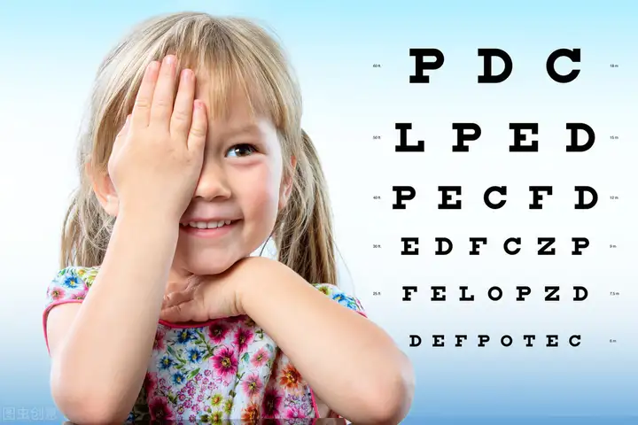
Vision test is to check the eyes’ ability to see objects at near and far distances. This Vision test is generally divide into two categories: far vision and near vision. It is tested with a vision chart. Normal near vision is to see 1.0 line clearly at 30 cm. The vision chart used in medical tests mainly checks central vision, that is, checks the visual acuity of the fovea of the retina, so that the initial situation of visual function can be easily and quickly understood.
2. Ophthalmoscopy.
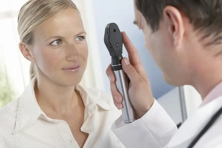
Fundus examination is an important method for examining vitreous, retinal, choroidal and optic nerve diseases. It is used to examine the refractive medium (cornea, aqueous humor, lens and vitreous) and fundus (optic disc, retina and choroid) of the eye, and is a common examination method in ophthalmology. The examination method is divided into direct examination method and indirect examination method, and is performed in a dark room.
3. Visual field test.
When the eyes are not moving and looking at a fixed object in front of the eyes, the range seen by the retinal photoreceptor cells outside the fovea of the macula is called the visual field. Visual field test methods are divided into dynamic and static tests. Visual field test is of great significance in the diagnosis of visual pathway diseases. For example, when the visual pathway is damaged by tumors in the sella turcica area or brain trauma, or when glaucoma or optic nerve atrophy occurs, different forms of visual field defects will appear.
4. Color vision test.
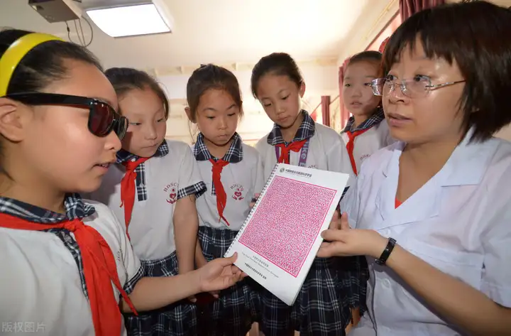
Color vision test is to test the color discrimination ability of the eyes. The human eye can generally distinguish 7 main colors including purple, blue, cyan, green, yellow, orange and red. The test method is to distinguish the color spectrum or pattern composed of various colors.
It is generally divided into four types: normal, color weak, monochrome discrimination (usually called color blindness, which means the inability to distinguish any color) and monochrome discrimination.
5. Dark adaptation test.
When a person goes from a bright place to a dark place, he can’t see anything in the first moment. Later, due to the resynthesis of rhodopsin in rod cells, the sensitivity of the retina to weak light gradually increases, and then he can see something. This process is called dark adaptation. People with impaired or impaired dark adaptation ability (night blindness) have very poor vision in weak light and difficulty in movement, which affects or even makes it impossible to work at night. Youneng Eyewash reminds you that clinically, vitamin A deficiency, glaucoma, and certain retinal and optic nerve diseases can reduce the sensitivity of retinal photosensitivity.
6. Stereoscopic vision test.
Stereoscopic vision, also known as stereoscopic acuity, refers to the human eye’s ability to perceive the front and back, distance, height, depth, distance, speed, etc. of objects in three-dimensional space. Poor stereoscopic vision will cause errors in judging the relative position of objects in space and poor tracking ability of moving objects, such as not seeing clearly when jumping up to grab the horizontal bar, or unclear judgment of the direction, distance, and speed of the ball when playing ball.
7. Contrast sensitivity test.
The lower the contrast threshold, the higher the contrast sensitivity, and the better the vision. It is also proportional to the degree of myopia. The lower the degree, the higher the sensitivity, and the higher the degree, the worse the sensitivity. The younger the age, the higher the contrast sensitivity. Testing the contrast sensitivity of amblyopic children and low vision patients is also of great significance for the comprehensive evaluation of visual quality.
8. Visual electrophysiological examination.
When the eyes are stimulated by light or graphics, they will produce tiny electrical activities such as potential and current. This is visual electrophysiology. The electrical activities of normal people and patients with eye diseases are different. Therefore, some eye diseases can be diagnosed through visual electrophysiological examination. The examination methods include electrooculogram, electroretinogram and visual evoked potential. Visual electrophysiological examination is not only suitable for general patients, but also for infants, mentally retarded people or pseudo-blind people.
9. Slit lamp microscopy.
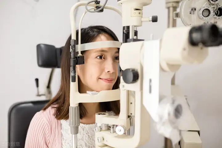
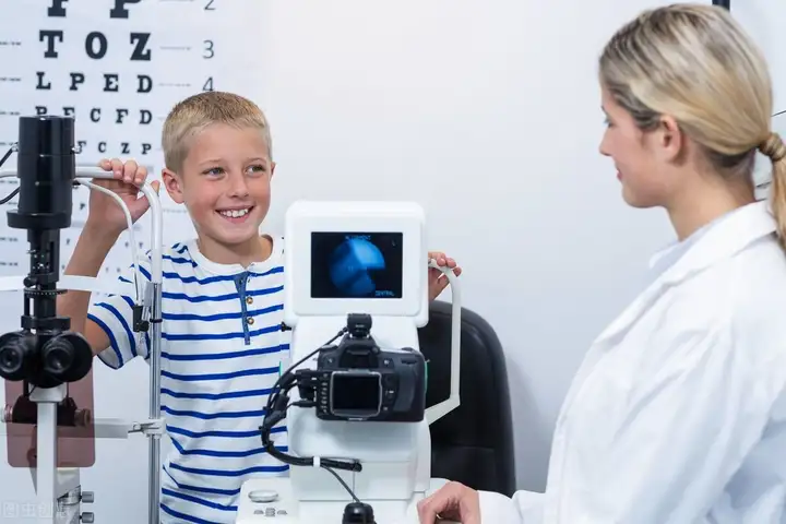
Slit lamp microscopy consists of a lighting system and a binocular microscope. It is a method of examining eye diseases in a dark room. The principle of use is that light passes through a slit to illuminate the eye. Slit lamp microscopy can clearly observe the lesions of the eyelids, conjunctiva, sclera, cornea, anterior chamber, iris, pupil lens and the front 1/3 of the vitreous body. The location, nature, size and depth of the lesions can be determined. It is an important instrument that is indispensable for ophthalmic examinations.
10. Gonioscopy.
The state of the anterior chamber angle is an important window to distinguish different types of glaucoma, and gonioscopy is the “key” to open this window. Gonioscopy is to check the width of the anterior chamber angle and its width changes when the intraocular pressure fluctuates. It is of great value in the diagnosis and treatment of various glaucoma. In addition, gonioscopy is also helpful for the diagnosis of foreign bodies in the anterior chamber angle or tumors at the root of the iris, new blood vessels, etc.
11. Intraocular pressure test.
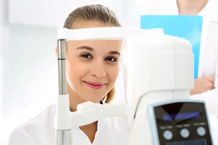
Intraocular pressure is the pressure inside the eyeball. The normal intraocular pressure of a person is 1.47-2.79 kPa (11-21 mmHg) to maintain the normal shape of the eyeball and the visual function of the eye. The main function of intraocular pressure is to maintain the shape of the eyeball and normal physiological function. It cannot be too high or too low. The method of detecting intraocular pressure refers to the use of medical equipment and medical methods to detect intraocular pressure. Through intraocular pressure detection, the patient’s condition such as glaucoma can be controlled, and eye diseases that may occur in patients with diabetes and hypertension can be effectively prevented. The main methods of detecting intraocular pressure are finger pressure measurement and tonometer measurement.
12. Mydriasis examination.
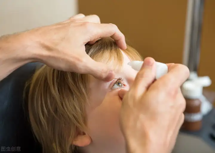
Mydriasis examination is to perform optometry after the ciliary muscles of the eyes are completely paralyz by drugs and lose their accommodative function. We reminds you that since the accommodative power of adolescent eyes is strong, it is difficult to rule out pseudomyopia if the pupils are not dilate during optometry. Therefore, children under 16 years old who are getting glasses for the first time and children with hyperopia must have their pupils dilated before optometry. The methods of mydriasis optometry for patients of different ages are different.
13. Optometry.
Optometry is to check the accuracy of the glasses you wear. There are two methods of optometry: subjective optometry and objective optometry. Subjective optometry is also call the lens insertion method or the obvious optometry method. The lens is place in front of the patient to perform a corrective vision test to determine the nature of the refractive error and the appropriate corrective lens degree. The most commonly used objective optometry methods are retinoscopy and computer optometry.
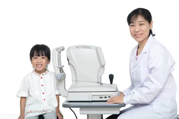
Eyes are a routine check-up item in the physical examination. Youneng Eye Wash recommends that normal people should have an eye check-up once a year. The items to be check include vision, eyelids, eyelashes, conjunctiva, pupils, fundus and intraocular pressure, etc. The doctor will perform different examinations according to the eye conditions of different people.

