

The information provided in the article “T1 vs T2 MRI: 5 Key Differences Explained” is intended for general informational and educational purposes only and does not constitute professional medical advice, diagnosis, or treatment. Always seek the advice of your physician or other qualified health provider with any questions you may have regarding a medical condition or interpreting diagnostic scans.
This article was written by Dr. Himanshu Gupta, Orthopedics & Robotics, Amicare Hospital.

🏥 Medical Review Board Member
Dr. Himanshu Gupta
Orthopedic Specialist | Amicare Hospital (Ghaziabad, India)
Dr. Himanshu Gupta is a distinguished medical professional with over 20 years of expertise in orthopedics and joint replacement surgery. As the Chief Orthopedic and Director at Amicare Hospital, he specializes in advanced robotic-assisted procedures and sports injury management, dedicated to enhancing mobility and quality of life for his patients. Dr. Gupta is committed to bridging the gap between complex surgical clinical practice and accessible, evidence-based patient education. His rigorous approach to medical oversight ensures that every piece of content he reviews meets the highest standards of clinical accuracy, safety, and reliability.
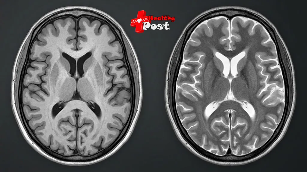
If you have ever compared two scans of the same brain and noticed that the fluid is black in one and glowing white in the other, you have already met the core idea behind t1 vs t2 mri. Both are ordinary MRI images, but each is tuned to emphasize a different tissue property. T1-weighted images are especially good at showing anatomy, with fat bright and fluid dark. T2-weighted images are especially good at revealing fluid and many abnormalities, with fluid bright.
Neither one is better than the other, which is why radiologists almost always read them together. This article explains what T1 and T2 mean, walks through the five main differences, shows how to tell the two image types apart, and covers FLAIR, STIR and contrast-enhanced scans along the way, including what a bright spot on a scan does and does not tell you.
How an MRI Creates a Picture
MRI uses a strong magnet, radiofrequency pulses and computer processing to build images of the inside of the body. The scanner mostly listens to hydrogen nuclei (protons), which are plentiful in water and fat. A radio pulse briefly knocks these protons out of alignment, and as they settle back they release signals that receiver coils pick up and software turns into pictures.
Because hydrogen in a pool of cerebrospinal fluid (CSF), a layer of fat and a patch of muscle sits in very different molecular surroundings, each gives off a different signal. An MRI sequence is a specific recipe of pulses, magnetic field gradients and timing designed to emphasize one kind of difference. A single exam usually strings several sequences together.
There is no ionizing radiation involved, which is one reason MRI is often chosen for soft tissue. If you are weighing it against another test, this overview of how MRI compares with CT lays out the main trade-offs.
What T1 and T2 Actually Measure
After a radio pulse, two things happen at once. Magnetization along the main magnetic field slowly rebuilds, and magnetization across that field fades as the protons drift out of step. These are T1 and T2 relaxation, and they are the raw material of MRI contrast. StatPearls’ MRI overview describes them as the relaxation times that govern image contrast.
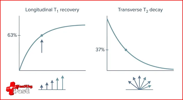
T1 is the longitudinal relaxation time, also called spin-lattice relaxation. It describes how long protons take to hand energy back to their surroundings and regain alignment with the main field. By convention it is the time needed for roughly 63% of that recovery. Fat has a relatively short T1, while free water has a long one.
T2 is the transverse relaxation time, or spin-spin relaxation. It describes how quickly protons lose phase coherence, the synchronized wobble that produces a measurable signal. It is the time for that signal to fall to about 37% of its starting value. Free water has a long T2, whereas tissues with more restricted molecular environments tend to have shorter ones.
Both are intrinsic tissue properties, although measured values shift with magnetic field strength, temperature and tissue composition. That is one reason a 3T scanner does not produce quite the same contrast as a 1.5T scanner.
What Does T2 Mean on an MRI?
When a report calls an image T2-weighted, the sequence was set up so that differences in T2 relaxation dominate the contrast. Tissue with a long T2, such as fluid, appears bright. A bright area is often described as T2 hyperintensity, and it usually reflects extra water: edema, inflammation, a cyst or certain tumors, for example. It is a clue rather than a verdict, because many normal structures are bright on T2 as well.
One related term causes frequent confusion. T2* (T2-star) also includes signal loss from magnetic field inconsistencies and susceptibility effects, so it is not the same thing as T2.
T1 vs T2 MRI: The 5 Key Differences
This is the short version of T1 weighted vs T2 weighted MRI. The paragraphs after the table explain each row.
| Feature | T1-weighted | T2-weighted |
|---|---|---|
| 1. Relaxation emphasized | Longitudinal recovery (T1) | Transverse decay (T2) |
| 2. Fluid, including CSF | Usually dark | Usually bright |
| 3. Fat | Bright | Often bright; depends on the sequence |
| 4. Main strength | Anatomy and tissue structure | Fluid-related change and many abnormalities |
| 5. Typical uses | Anatomy, bone marrow, post-contrast scans | Edema, inflammation, many lesions |
1. What each one emphasizes. T1 weighting highlights how quickly longitudinal magnetization recovers, and T2 weighting highlights how quickly the transverse signal decays. This is the technical root of every other difference, and it is controlled by the timing settings described later.
2. How fluid looks. This is the easiest of the T1 and T2 MRI differences for most people to remember. On conventional T1-weighted images, CSF and other free fluid are dark. On conventional T2-weighted images, they are bright. There are exceptions, since fluid that contains protein or blood products can look different, and subacute bleeding is relatively bright on T1.
3. How fat looks. Fat is bright on T1. On T2 the answer depends on technique. On conventional fast spin-echo T2 images fat often stays fairly bright, and it turns dark mainly when fat suppression is applied. So the rule of thumb that fat is dark on T2 is not reliable.
4. What each is best at. T1 gives a clean anatomical map. In the brain, white matter looks brighter than gray matter on T1 and the reverse on T2, partly because gray matter contains somewhat more water. Clinical references describe T2-weighted images as more sensitive to demyelination, infarction, edema and chronic hemorrhage, while T1-weighted images are more sensitive to fat and subacute hemorrhage.
5. Where each is used. Both appear in brain, spine, joint, abdominal and pelvic examinations, but the balance shifts with the body part and the question being asked. Spine protocols typically include both T1 and T2 slices, often alongside a STIR sequence. A knee exam leans on fluid-sensitive images to look at cartilage, ligaments and joint effusions. For background on the joint and spine problems that commonly lead to an MRI, this guide to common orthopedic conditions is a useful starting point.
T1 MRI vs T2 MRI Images: How to Tell Them Apart
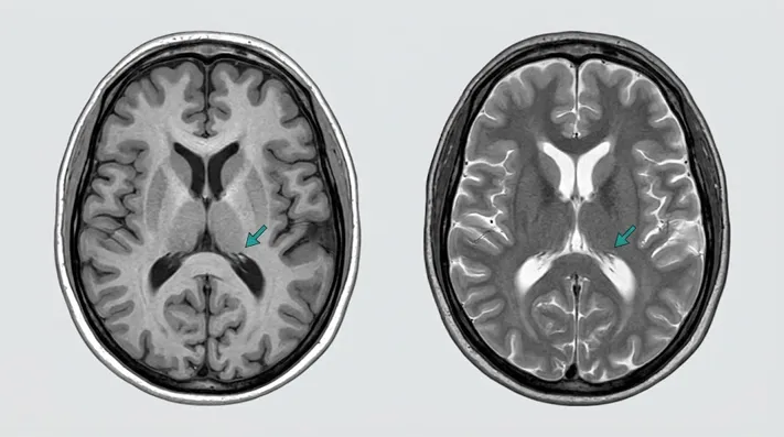
If you are handed two unlabeled brain scans, the CSF is the fastest clue.
On a T1-weighted image, look for:
- dark CSF in the ventricles
- white matter that looks brighter than gray matter
- bright fat, such as the layer beneath the scalp
On a T2-weighted image, look for:
- bright CSF in the ventricles and around the brain
- gray matter that looks slightly brighter than white matter
- fluid-containing structures that appear bright
On both, air and cortical bone look dark, because they contain few mobile hydrogen protons to generate signal. That is also why bone injuries are usually assessed differently; this explainer on how bone fractures appear on an X-ray shows what radiologists look for on a plain film.
A few things can mislead you. FLAIR images are built on T2 contrast but suppress CSF, so the ventricles look dark even though the tissue contrast resembles T2. Fat-suppressed scans turn normally bright fat dark. And T1 images taken after contrast show bright enhancing areas that were not bright before. When it matters, check the sequence label rather than relying on appearance alone.
Why Doctors Order More Than One Sequence
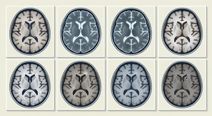
No single sequence answers every question. A brain MRI might combine several, each contributing something different:
T1-weighted imaging shows anatomy and tissue architecture.
T2-weighted imaging highlights fluid and abnormal water content.
FLAIR stands for Fluid-Attenuated Inversion Recovery. It suppresses the bright signal from normal CSF, which makes abnormalities near the ventricles and other fluid spaces easier to see. It is widely used for conditions such as multiple sclerosis, some infarctions, white matter abnormalities and inflammation, although it cannot name the cause of a finding on its own.
STIR (Short Tau Inversion Recovery) suppresses signal from fat. It helps reveal bone marrow edema, muscle and soft-tissue injuries, and inflammation in the spine and joints that bright fat might otherwise hide. FLAIR and STIR use similar inversion-recovery principles but cancel out different tissues.
Diffusion-weighted imaging and proton density imaging add further information, and contrast-enhanced T1 is covered below.
Think of a lesion in the brain. T1 shows where it sits, T2 shows the water-rich area in and around it, FLAIR clarifies surrounding tissue changes, and post-contrast T1 shows where enhancement occurs. The bright halo on T2 or FLAIR might be edema, infiltrating tumor, inflammation or something else, and radiologists weigh all of it together with the patient’s symptoms.
TR, TE and TI: The Timing Behind the Contrast
MRI technologists create T1 or T2 weighting mainly by adjusting timing.
- TR (repetition time) is the interval between successive excitation pulses. A short TR increases T1 weighting.
- TE (echo time) is the time between the excitation pulse and the peak of the measured echo. A long TE increases T2 weighting.
- TI (inversion time) is the delay between an inversion pulse and the following excitation pulse. It is central to FLAIR and STIR.
| Image type | Typical timing approach |
|---|---|
| T1-weighted | Short TR, short TE |
| T2-weighted | Long TR, long TE |
| Proton density | Long TR, short TE |
| FLAIR | Inversion recovery tuned to cancel CSF |
| STIR | Inversion recovery tuned to cancel fat |
These are general principles rather than fixed numbers. Actual values depend on the scanner, field strength, pulse sequence and clinical question.
What Contrast-Enhanced T1 Adds
Contrast-enhanced T1 imaging is performed after an injected contrast agent, most commonly a gadolinium-based one, when it is clinically appropriate. These agents shorten the T1 of nearby tissue, so enhancing areas become brighter on T1-weighted images, while the effect on T2-weighted images is generally negligible. That is why enhancement is judged by comparing T1 images before and after the injection. The StatPearls review of gadolinium-based MRI contrast agents explains the mechanism in more detail.
Doctors use it to evaluate tumors, infections, inflammatory disease and disruption of the blood-brain barrier, among other things. Enhancement reflects both blood supply and how permeable the vessels are, so it does not simply mean that a lesion has a rich blood supply. Not every tumor enhances, and enhancement alone cannot establish a specific diagnosis.
Before contrast, tell the MRI team about kidney or liver disease, pregnancy or breastfeeding. Nephrogenic systemic fibrosis is a rare but serious complication that occurs almost exclusively in people with significantly impaired kidney function, and newer macrocyclic agents are considered more stable and carry a lower risk.
Can T1 or T2 MRI Detect Cancer?
Both sequences can help detect and characterize abnormalities, including tumors, but the signal pattern alone does not decide whether something is cancer. A bright T2 signal can be edema, inflammation, a cyst or a normal structure. A dark T1 signal is just as non-specific. Some tumors have mixed signal, some benign lesions resemble malignant ones, and contrast enhancement is not exclusive to cancer.
A radiologist interprets findings across all sequences, alongside clinical information. A definitive cancer diagnosis may require additional imaging, specialist assessment or tissue sampling. If your report mentions a T2 hyperintensity and you are worried, ask the doctor who requested the scan what it means for you specifically.
Frequently Asked Questions
Why is fluid dark on T1 but bright on T2? Fluid has long T1 and T2 times. With the short timing used for T1 weighting, little of its longitudinal magnetization has recovered, so it gives a weak signal. With the long timing used for T2 weighting, its signal persists, so it looks bright.
Is FLAIR a T1 or a T2 sequence? FLAIR is generally described as T2-based, with the CSF signal suppressed by an inversion pulse. Tissue contrast resembles T2, but free fluid appears dark.
Does every MRI include contrast? No. Many scans are done without it. Whether contrast is needed depends on the condition being investigated, and the decision is made by the referring clinician and radiologist.
Should a T2 hyperintensity on my report worry me? It means an area is brighter than expected on a T2-weighted image, usually because of more water. Causes range from harmless to significant, so the finding needs interpretation in context rather than alarm.
Do T1 and T2 images look the same on 1.5T and 3T scanners? The principles are the same, but tissue relaxation times change with field strength, so settings are adjusted. The images may differ slightly in contrast and detail.
Conclusion
The simplest way to remember t1 vs t2 mri is to look at the fluid: dark on T1, bright on T2. Behind that rule is a useful division of labor, with T1 describing structure and T2 revealing fluid-related change, and with FLAIR, STIR and contrast adding detail where needed. Neither sequence is universally better, and a radiologist’s report draws on all of them together.
If you have an MRI coming up or a report you do not fully understand, bring your questions to the doctor who ordered it. Knowing the basics of how these images are built makes that conversation easier.
References
- StatPearls. MRI Patient Safety And Care. NCBI Bookshelf, National Library of Medicine.
- Ibrahim MA, Hazhirkarzar B, Dublin AB. Gadolinium-Based MRI Contrast Agents. StatPearls, NCBI Bookshelf, National Library of Medicine.