
Metacarpal fracture postoperative rehabilitation
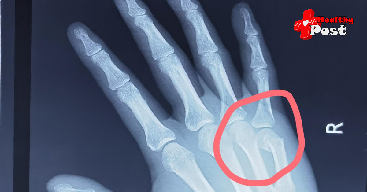
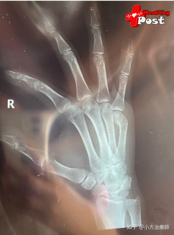
Metacarpal fracture is a common hand fracture in clinical practice. People cannot work without hands, which makes hands vulnerable to injury. Hands are fine motor organs of the human body. After a fracture occurs, it must be treated systematically in time, otherwise it is easy to affect the recovery of the fracture. If it is not treated in time and reasonably, it will cause the patient’s hand function to be impaired, affecting the quality of life in the future.
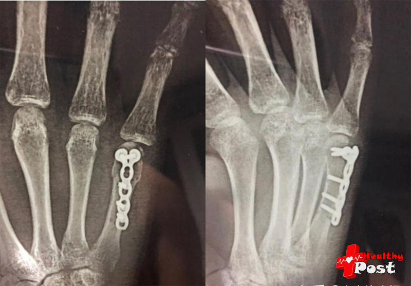
There are many treatment options. Surgical treatment has a significant effect in the treatment of metacarpal fractures, and postoperative rehabilitation is an important part of the recovery process.
Postoperative recovery can be divided into three stages:
Phase 1: Inflammation/Protection
Time: Week 1
Purpose: Protective immobilization, edema control, wound healing, full range of motion of uninvolved upper extremity joints
suggestion:
Edema control
- Elevate the affected limb
2. Cold wrap, mild retrograde massage, and mild pressure bandage if the fracture is stable
3. Active range of motion training of unaffected joints
4. Reshape the splint after edema subsides to maintain proper fit and positioning
Active/assisted active range of motion (A/AAROM) of uninvolved joints
1. Move all upper limb joints, except those adjacent to the fracture site
2. Avoid using elastic gloves to avoid applying uncontrollable external forces to the fracture when putting on and taking off
3. Do not move the joints adjacent to the fracture site until the fracture is stable after surgical fixation or clinical healing.
Phase II: Stabilization
Time: Week 2 to Week 6
Purpose: Control edema, no adhesion of scar, free sliding of flexor and extensor tendons, functional activities of joints adjacent to fracture
suggestion:
- Edema control: After the wound has healed and the fracture has stabilized, alternating hot and cold baths and elastic gloves can be added
2. Scar treatment: scar massage, silicone scar pad
3. Heat to prepare soft tissue for ROM and scar management
4. A/AAROM of the joints adjacent to the fracture, sliding and blocking exercises of the flexor and extensor tendons, and unresisted exercises or activities (avoiding additional passive pressure on the fracture site)
5. Perform mild functional activities of the hand under external fixation, and gradually progress to guided mild activities without external fixation. 6. In addition to personal hygiene and exercise activities, the brace should continue to be used for fixation and protection.
Stage 3: Fracture healing
Time: Week 7-10
Objective: To achieve full range or maximum range of motion with free tendon gliding, to have the muscle strength and endurance required for functional functions, and to be able to perform activities of daily living (ADL) independently
suggestion:
- Except for protective splints under the guidance of a physician
2. Continue edema and scar control
3. Perform thermal therapy (including paraffin and ultrasound thermal therapy ) to prepare soft tissue for ROM, joint movement, and scar mobilization
4. Active range of motion exercises (AROM), including flexor and extensor exercises, tendon gliding and blocking exercises; add blocking splints when necessary to avoid excessive dorsiflexion of the adhesion tendon
5. Treat joint stiffness through passive range of motion (PROM), stretching, and joint mobilization
6. Treat persistent joints with sequential static and static progressive splinting. Do not use antagonism, dorsiflexion, or static progressive splinting until the fracture is adequately healed.
7. Resistance exercises for the intrinsic and extrinsic muscles of the wrist and fingers
8. Perform functional and adaptive activities to achieve self-care in ADLs
The need for postoperative rehabilitation
It is very necessary to do rehabilitation exercises for the affected hand after surgery, mainly because the motor function of the hand is very complex and delicate.
The purpose of post-fracture rehabilitation is to reduce tissue adhesion around the fracture, restore joint range of motion and muscle strength. Early rehabilitation is aimed at restoring joint range of motion . Rehabilitation can also improve blood circulation in the affected hand , eliminate vascular spasms, prevent blood vessel adhesion and joint stiffness, and promote functional recovery, thereby accelerating fracture healing . Improving joint range of motion and softening scar tissue are also important for solving the problem of limited flexion and extension of the affected hand.
Muscle training
Muscle contraction is divided into static contraction and dynamic contraction.
Static contraction can promote venous and lymphatic return and accelerate the absorption of exudates; dynamic contraction can not only promote venous return, but also prevent adhesion and stiffness of muscles, tendons and joints in the early stage. Dynamic contraction of the hand is to perform muscle strength exercises. Muscle strength exercises must gradually increase strength according to the degree of wound surface and wound bleeding, the disappearance of tissue edema, and wound healing. Resistance exercises are not recommended before fracture healing.
Physical therapy
The application of physical therapy is mainly to improve blood circulation and promote the disappearance of edema.
Physical therapy can also promote the absorption and dissipation of inflammation, relieve pain in nerves, joints, and muscles, prevent muscle atrophy, local sensory disorders, etc., and help loosen adhesions and soften scars in the later stages.
The basic purpose of active joint exercise is to prevent adhesion and contracture of muscles, tendons and joint capsules. The premise of joint activity exercise is the guarantee of strong internal fixation of the hand. The joint movement should be performed one by one, and the movement should be smooth and slow. With appropriate strength, the maximum amplitude should be achieved as much as possible, and the force should be applied to the extent that it causes pain. When performing passive exercise on patients, the degree of force must be carefully controlled according to the patient’s pain sensation to avoid causing severe pain, and violence should not be applied to avoid causing new injuries.