


🏥 Medical Review Board Member
Dr. Himanshu Gupta
Orthopedic Specialist | Amicare Hospital (Ghaziabad, India)
Dr. Himanshu Gupta is a distinguished medical professional with over 20 years of expertise in orthopedics and joint replacement surgery. As the Chief Orthopedic and Director at Amicare Hospital, he specializes in advanced robotic-assisted procedures and sports injury management, dedicated to enhancing mobility and quality of life for his patients. Dr. Gupta is committed to bridging the gap between complex surgical clinical practice and accessible, evidence-based patient education. His rigorous approach to medical oversight ensures that every piece of content he reviews meets the highest standards of clinical accuracy, safety, and reliability.
Seeing an X-ray of your own injured wrist, ankle or arm can feel strangely disorienting. The image looks like a ghostly negative, the report seems written in code, and one question dominates: is it broken?
Knowing how to identify a fracture on an X-ray comes down to four clues. They are a break or step in the bone’s bright outer edge, a dark line (or sometimes an unusually bright band) running through the bone, bone ends that no longer line up, and indirect signs such as swelling in the surrounding tissue. This article explains each one in plain language, shows how normal and fractured bones differ, and decodes the terms you’ll meet in a radiology report.
One caution first. These clues help you follow the conversation with your care team. They don’t replace it. Some fractures are subtle, and some don’t show on the first X-ray at all. Reliable interpretation needs trained eyes that read the images alongside your symptoms, the examination and how the injury happened.
What does a bone fracture look like on an X-ray?
X-rays pass a small dose of radiation through the body. Dense tissue such as bone absorbs more of it and appears white, softer tissue appears grey, and air looks nearly black. A healthy long bone, like the forearm or shin, shows up as a bright, sharply outlined shaft with a greyer, lacy interior.
A fracture disrupts that orderly picture. You might see a thin dark gap where the bone has separated, a crumpled area that looks denser than it should, or an outline that bends where it should run straight. An X-ray also flattens a three-dimensional bone into a two-dimensional shadow, so a crack that is obvious from the front can nearly vanish from the side. That’s why clinicians usually request at least two views from different angles.
Treat the four clues below as a vocabulary, not a checklist. A fracture may show one clue, several, or, early on, none.
Four key clues to look for on a bone X-ray
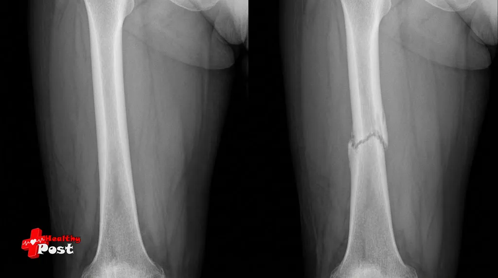
Clue 1: Signs of cortical disruption on an X-ray
The cortex is the dense outer shell of the bone. On an X-ray it forms a bright, continuous line along each edge of the shaft, a bit like the outline of a drawing. In a healthy bone it’s smooth and unbroken, so an imaginary finger traced around it wouldn’t catch.
Signs of cortical disruption on an X-ray include a visible gap, a small step where one side sits out of line with the other, a sharp notch or angle, and a localised bulge. In children, whose bones are softer, a “buckle” (torus) fracture often appears as a gentle ripple in the cortex rather than a clean break.
Cortical changes are among the strongest clues, but they aren’t foolproof. Tiny channels that carry blood vessels into the bone, overlapping structures and skin folds can mimic a break. Meanwhile, early stress fractures and some impacted fractures can leave the cortex looking almost normal. A subtle irregularity is a reason to look harder, not a verdict.
Clue 2: A visible fracture line or abnormal bone density
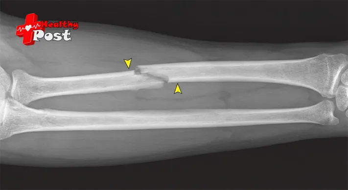
The most familiar sign is a dark line crossing the bone, called a lucent or fracture line. It marks where the bone has separated and less material blocks the beam. Its direction hints at the injury. A transverse line runs across the bone, often after a direct blow or bending force. An oblique line runs diagonally. A spiral line wraps around the shaft after a twisting injury. In a comminuted fracture the bone has split into several pieces, so multiple lines may radiate from one point.
Not every fracture looks darker, though. When bone ends are driven into each other, the overlap can look brighter or denser than the bone around it, which is typical of impacted fractures. In the spine, a collapsed vertebra may look flattened or wedge-shaped rather than cracked, which is how compression fractures of the spine often appear on imaging.
Many harmless features imitate a fracture line. Growth plates in children, accessory bones, the normal seams where bones meet, and even a fold of clothing can resemble a crack. Interpreting a possible fracture line takes anatomical knowledge as well as a sharp eye.
Clue 3: Abnormal bone alignment or displaced fragments
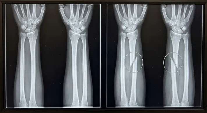
Bones have an expected shape and axis, and a break can change that geometry. Fragments may shift sideways, tilt at an angle (angulation), rotate, overlap, or leave the limb shortened. Any of these suggests a displaced fracture. Radiologists compare the bone’s overall line on each view and, when in doubt, with the opposite limb.
Displacement shapes treatment. A nondisplaced fracture, where the ends stay close to their normal position, can often be supported with a cast or splint. A significantly displaced one may need realignment, sometimes with surgery. The reverse reading isn’t safe, however: normal-looking alignment does not rule out a fracture, because a nondisplaced break can look almost undisturbed.
Joint surfaces deserve special attention. When a break extends into a joint, even a small step or depressed fragment can affect long-term movement. A tibial plateau fracture, at the top of the shin bone near the knee, shows why clinicians study these surfaces closely and sometimes request a CT scan for a clearer view.
Clue 4: Indirect signs of bone injury
Sometimes the bone itself looks unremarkable and the clue lies around it. Soft-tissue swelling appears as a bulge or a blurring of the normally crisp planes of fat and muscle beside the bone. Swelling alone doesn’t prove a break, since sprains and bruises cause it too, but swelling directly over a tender bone raises suspicion.
Joint fluid can be revealing. In the elbow, an effusion pushes small pads of fat away from the bone, producing a dark triangle known as the fat pad sign. It can point to a fracture hidden within the joint, particularly in children. In the knee, a layer of fat floating on blood inside the joint suggests a break reaching the joint surface.
Other clues appear as healing begins. A thin line of new bone along the shaft (periosteal reaction) or a hazy cloud of callus around the break can show up after the first week or two. That’s one reason a repeat X-ray sometimes reveals a fracture that was invisible at first. The stages of fracture healing, from the initial blood clot through to remodelling over months, explain what these later changes represent.
None of these signs proves a fracture alone. Clinicians weigh them against where it hurts, what the examination shows and whether other views agree. If symptoms strongly suggest a fracture despite normal images, further assessment may be needed.
Normal vs. fractured bone X-ray: what changes
Anyone learning how to identify a fracture on an X-ray benefits from a baseline. Treat the normal bone as the reference and look for departures from it.
| Feature | Normal bone | Possible fracture |
|---|---|---|
| Outer cortex | Smooth, continuous | Break, step or ripple |
| Bone interior | Even density, lacy texture | Dark line or denser band |
| Alignment | Expected position and axis | Angled, shifted, rotated or shortened |
| Contour | Intact outline | Deformed or compressed |
| Nearby tissue | Crisp tissue planes | Swelling or joint fluid |
These are general patterns, not absolute rules. Healthy anatomy can surprise you. Growth plates in children appear as dark bands near the ends of bones and are often mistaken for fractures. For that reason radiologists may take an X-ray of the unaffected limb for comparison, a practice RadiologyInfo.org describes in its bone X-ray guide. A nondisplaced or occult fracture may also produce no obvious abnormality on the first image.
How to read an X-ray report
A radiology report is a written summary by a radiologist, a doctor who specialises in medical imaging, and it’s usually sent to the clinician who requested the X-ray. Most reports follow a similar outline. The examination section names the body part and views. The clinical history states why the X-ray was requested. The findings describe what the radiologist sees, structure by structure, and the impression condenses the key points into a few lines. If you only read one part, start with the impression, then return to the findings for detail.
Common terms include:
- Nondisplaced fracture: a break whose fragments remain essentially in position.
- Displaced fracture: the fragments have moved out of their expected position.
- Transverse, oblique and comminuted fractures: breaks running across the bone, diagonally, or splitting it into multiple fragments.
- Angulation: the fragments meet at an angle instead of a straight line.
- Soft-tissue swelling: swelling around the injury, which can occur with or without a fracture.
- No acute osseous abnormality: no obvious recent bone injury is seen. This doesn’t always exclude an occult (hidden) fracture.
- “Cannot exclude” or “clinical correlation recommended”: the radiologist is flagging uncertainty and asking the treating clinician to match the images with the examination.
Phrases like “cannot exclude a nondisplaced fracture” deserve attention rather than alarm. They usually mean the image is suspicious or equivocal, and your clinician will decide whether to immobilise the area, repeat the X-ray or request another scan. If a report describes a displaced or unstable break, treatment may involve realignment and, in some cases, fixation with plates and screws.
Rather than interpreting a report alone, ask your treating clinician to walk you through it, especially when the findings are uncertain or symptoms persist. A radiologist’s report is always read alongside your history and examination.
Can an X-ray miss a fracture?
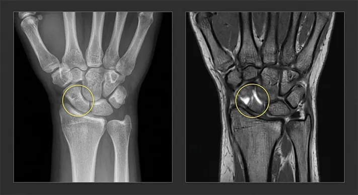
Yes. An initial X-ray can look normal even when a bone is broken. Hairline cracks, early stress fractures and breaks hidden behind overlapping bones can slip past the first image. The scaphoid, a small bone on the thumb side of the wrist, is the classic example. Hip fractures in older adults and rib fractures can also be difficult to see.
If tenderness, swelling or trouble bearing weight continues after a clear X-ray, clinicians may treat the injury as a possible fracture with a splint or restricted activity, then arrange a repeat X-ray after several days or a different kind of scan. RadiologyInfo notes that MRI can detect subtle or occult fractures and bone bruises not visible on X-ray, while CT can show complicated or subtle fractures in detail. For suspected scaphoid fractures specifically, the NICE guideline on non-complex fractures advises considering MRI as first-line imaging.
Persistent pain after a chest injury shows how a doubtful image and lasting symptoms can coexist. A rib fracture can be hard to confirm on a plain X-ray, yet pain and healing can run on for weeks.
When to seek medical attention
A suspected fracture should be assessed by a healthcare professional. MedlinePlus lists intense pain, deformity, swelling, bruising or tenderness, numbness and tingling, and problems moving a limb among the symptoms, and advises getting medical care right away.
Go to an emergency department (A&E in the UK) after a significant injury, or if there is severe pain, a visible deformity or you can’t use the limb. Call emergency services for bone protruding through the skin, uncontrolled bleeding, numbness, a limb that turns cold, pale or blue, or a suspected injury to the head, neck, back, hip or pelvis. Don’t rely on your own reading of an X-ray to rule out a fracture or delay care.
Frequently asked questions
Can I tell if my bone is broken just by looking at my own X-ray?
You can learn to spot obvious signs, such as a dark line or a step in the outline, but you can’t reliably diagnose a fracture this way. Normal structures can mimic breaks, and real breaks can be subtle. Interpretation requires clinical context.
How long after an injury does a hidden fracture show up?
It varies by bone and injury. As the body begins repairing a break, changes at the fracture edges can make it easier to see, which is why some pathways for suspected wrist (scaphoid) injuries include a repeat X-ray roughly 10 to 14 days later. Your clinician will decide on timing.
Why did the radiographer take several views, or X-ray my other limb?
A single view can hide a fracture that lies edge-on to the beam, so extra angles help. Images of the unaffected limb provide a personal baseline, which is especially useful for children’s growth plates.
Is a hairline fracture less serious than a full break?
Not necessarily. A hairline fracture is a thin crack that may not separate the bone, but its seriousness depends on location, cause and whether it affects a joint or weight-bearing bone. It still needs professional assessment.
Is the radiation from a fracture X-ray a concern?
A bone X-ray uses a very small dose of radiation, and RadiologyInfo states that the benefit of an accurate diagnosis far outweighs the risk. Tell the technologist if there’s any chance you might be pregnant.
Putting the four clues together
Knowing how to identify a fracture on an X-ray is mostly about pattern recognition: a cortex that’s no longer smooth, a line or density that shouldn’t be there, alignment that has shifted, and swelling or fluid that hints at injury underneath. Comparing normal and fractured bone makes those departures easier to see, and understanding report terms like displaced, nondisplaced and “no acute osseous abnormality” makes the results less intimidating.
The most useful next step is a conversation. Bring your questions, and your symptoms, to the clinician treating you. If pain persists after a normal-looking X-ray, say so, because a repeat X-ray or an MRI or CT may be the right answer.
References
- RadiologyInfo.org (RSNA/ACR), “Bone X-ray (Radiography)”: https://www.radiologyinfo.org/en/info/bonerad
- MedlinePlus (U.S. National Library of Medicine), “Fractures”: https://medlineplus.gov/fractures.html
- NICE, “Fractures (non-complex): assessment and management” (NG38): https://www.nice.org.uk/guidance/ng38