Let’s know ICL Surgery Introduction
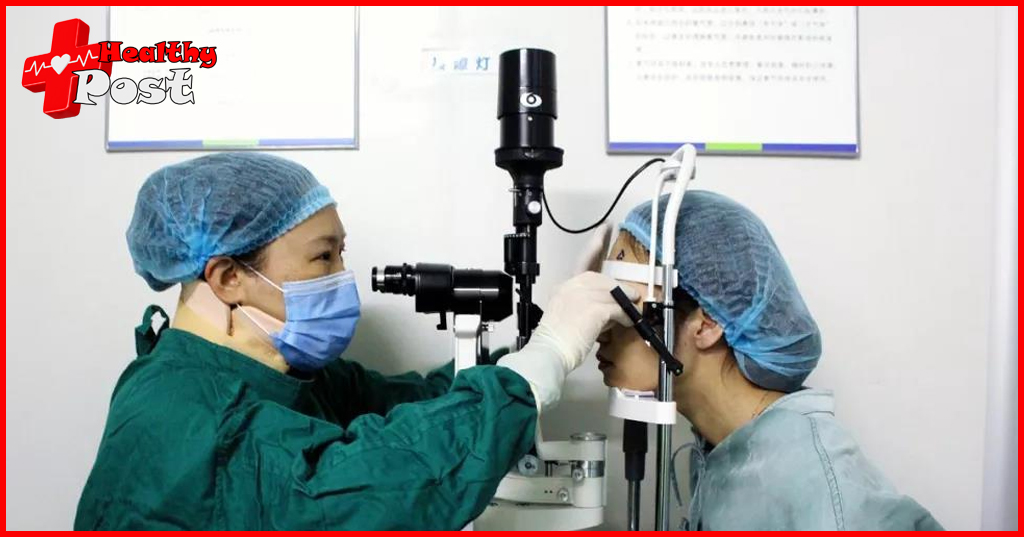
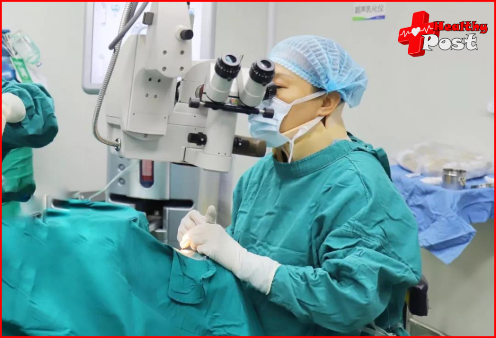
ICL surgery (implantable collamer lens) is a type of posterior chamber phakic intraocular lens implantation surgery, which is an “additive correction”. Long-term clinical trials have shown reliable safety and stable refractive correction effects, and have become one of the new research directions of refractive surgery . China began to introduce research and clinical trials of ICL surgery in 2006.
The surgical principle of posterior chamber ICL implantation is to implant an artificial lens with refractive power in the center of the posterior chamber, with both sides supported and fixed on the ciliary sulcus to correct refractive errors .
2. People who are suitable for ICL surgery:
1..21 years old – 45 years old.
2. If the increase in the number of degrees does not exceed 50 in a year, it is considered relatively stable.
3. Myopia 50 degrees – 2000 degrees, astigmatism less than 500 degrees, hyperopia 200 degrees – 1000 degrees.
4. Routine examination showed no abnormalities in the cornea, angle structure, intraocular pressure, or fundus.
5. The corneal thickness is not suitable for laser surgery . Ps: The best arch height after ICL surgery is 0.5-1.5 corneal thickness.
6. Anterior chamber (ACD) depth greater than 2.8 mm
The depth of the anterior chamber is the size of the space for your lens. It cannot be said that the bigger the better. The right distance is the best.
(1 millimeter = 1000 micrometers, that is, 1mm = 1000μm)
7. Corneal endothelial cell count greater than 2500/mm
The normal value of corneal endothelial cells varies according to age.
In normal people before the age of 30, the average corneal endothelial cell density is 3000-4000 cells/mm², about 3000 cells/mm² between 31-40 years old, about 2800 cells/mm² between 40-50 years old, about 2600 cells/mm² between 51-60 years old, and 2160-2400 cells/mm² between 61-80 years old.
Note: Long-term wearing of contact lenses can damage endothelial cells.
8. Dark pupil size is less than 6.5mm
If the dark pupil is relatively large, there will be glare and double vision. The average pupil size is about 2.5-4mm. In dark light conditions, the pupil will naturally dilate. This is called the dark pupil. Generally, the diameter of the dark pupil is less than 7mm. If it exceeds this value, the pupil is enlarged.
Bright pupil: photopic; dark pupil: mesopic
9. Corneal diameter (WTW) ≥10.8mm.
10. Those who are not mentally ill and have a reasonable desire to remove their glasses and appropriate postoperative expectations.

3. Not suitable for ICL
1. Active inflammation and tumors of the eyes and ocular appendages.
2. Corneal degeneration or corneal endothelial cell count < 2000/mm.
3. Corneal diameter (WTW) <10.5mm.
4. Anterior depth < 2.8mm.
5. Lens opacity that has significantly affected vision . Clearly diagnosed glaucoma; severe fundus lesions .
6. Huge irido-ciliary cyst at the loop position.
7. Pupil diameter > 7mm or pupil deviation in dim light.
8. People with mental disorders who have not undergone surgery with the permission of a psychologist or psychiatrist, women who are pregnant or breastfeeding, people with systemic collagen sensitivity or autoimmune diseases, and people with insulin-dependent diabetes.
9. The patient cannot understand the risks of surgery and is overly anxious.
10. Unstable degree.
4. Introduction of iCl crystal
1. Materials
ICL material is highly biocompatible hydrophilic collagen (-0.198), hydroxyethyl methacrylate (HEMA), referred to as collagen polymer. The most significant feature of this material is that it can naturally deposit a layer of fibrin on the surface of ICL , and fibrin can inhibit the integration of protein aqueous solution. The optical part of ICL is 49 ~ 5.8mm in diameter, the lens is ultra-thin, the thinnest part is only 50μm, the optical properties are close to the natural lens, it can absorb tight external rays, it can be folded and removed.
2. Specifications
The development of ICL lenses has gone through the upgrades of V1, V2, V3, V4 and V4c. The ICL product currently in clinical use is ICL-V4c. ICL-V4c has 4 lens diameters, namely 12.1mm, 12.6mm, 13.2mm and 13.7mm. Compared with ICL-V4, the correction range of the new ICL-V4ec starts from 50° myopia, the correction range of astigmatism is 50° ~ 600°, and mixed astigmatism can also be corrected. ICL-V4c has 3 new small holes in its design compared to ICL-V4, which allows the free flow of aqueous humor. It does not require preoperative iris laser circumcision, and will not significantly affect the optical quality and intraocular scattering after iris ligation.
The ICL-V4c lens has a hole. The central hole has the following functions: First, it can effectively allow the aqueous humor to flow continuously from the posterior chamber to the anterior chamber, effectively preventing postoperative high intraocular pressure caused by obstruction or too small iris periosteum. Second, the presence of the central hole may protect the lens by providing more natural aqueous humor flow, thereby reducing the occurrence of cataracts.
3.V5? V6?
V5 eliminates the process of taking the lens out of the bottle and pressing it on the injector. At the same time, the aperture area of V5 will be larger, so the aperture and glare problems will be improved to a certain extent, and the vision in low-light conditions will be better. It is suitable for vision below 1400 degrees.
Some people think that V5 has not undergone major changes compared to V4, so there is no need to wait for V4, and the price of V5 is much higher than that of V4.
V6 is still under development and is aimed at people with presbyopia.
5. Benefits of iCl
1. The surgical process is safer. ICL lens implantation does not require corneal cutting, but instead achieves the goal of restoring high-definition vision by implanting a micro-intraocular lens. It ensures the biomechanical stability of the cornea and the overall safety of the eye, with a shorter surgical time and faster postoperative recovery. (In short, the risks of ICL are mainly after the operation, while the risks of laser surgery are mainly during the operation.)
2. A wider correction range. It can correct myopia within 1800/2000 degrees and astigmatism within 500/600 degrees. It is especially suitable for people who are not suitable for myopia laser surgery due to insufficient corneal thickness, too high degree, dry eyes, etc.
3. Reversible. If the patient’s vision changes significantly and the ICL is no longer suitable, the ICL can be removed or replaced at any time.
4. Long-term use. Once an ICL lens is implanted, it will not be rejected by the human body and will not deteriorate or wear out.
6. Risks and sequelae of ICL
1. Intraocular infection
After all, it is an intraocular surgery. The risk of intraocular infection is definitely there (as long as the operation is disinfected, eye drops are used early after the operation, and no sewage is taken in, the probability is very low. You can refer to the 0.057% probability of endophthalmitis after cataract surgery ). Of course, if an intraocular infection occurs, it can be catastrophic.
At the same time, sensitivity to strong light, immunity to anesthetics, too little anesthetics, loss of anesthetic effectiveness, sensitive pain nerves , etc. during the operation may cause the eyes to move unconsciously during the operation, resulting in larger surgical incisions, poor incision positions, and excessive bleeding. However, the blood halo in the whites of the eyes generally subsides after 1 to 2 weeks.

2. Glare, aperture, ghosting
The pupil is a small round hole in the center of the iris in an animal or human eye, which is the channel for light to enter the eye. The contraction of the pupillary sphincter on the iris can shrink the pupil, and the contraction of the pupil dilator muscle can dilate the pupil. The dilation and contraction of the pupil control the amount of light entering the pupil. When encountering strong light stimulation, the pupil will shrink, and in a dark room, the pupil will naturally dilate.
The normal pupil diameter is 2.5~4mm, with a difference of less than 0.25mm between the two eyes. In a dark room, the pupil diameter is 5~7mm. That is, the general dark pupil diameter is less than 7mm. If it exceeds this value, the pupil is enlarged. The pupil size of the two eyes of normal people should be about the same.
Glare and ghosting?
A dark pupil that is too large will increase the reaction of postoperative glare , aperture, etc., which is similar to the effect of astigmatism, especially when looking at luminous objects. Most people will adapt or improve this reaction over time.
However, pupil size is innate, and there is currently no treatment for the sequelae of glare and double vision caused by large dark pupils. Almost all refractive surgeries may cause glare, so preoperative screening should be done and carefully considered. If the dark pupil is too large and you are worried about glare after surgery, you can first consider wearing frame lenses or contact lenses to correct your vision.
3. Vault height and glaucoma and cataract
Vault is a key factor in determining the success or failure of ICL surgery. The distance between the ICL and the lens is usually expressed by the vault (the distance between the posterior surface of the ICL and the anterior capsule of the lens). The vault is determined by the natural curvature of the ICL, the length of the ICL and the length of the ciliary sulcus (anterior chamber depth = vault + implanted lens thickness + residual anterior chamber depth). Clinically, the length of the ICL is generally estimated by measuring the transverse corneal diameter (WTW distance from 3 o’clock to 9 o’clock) and adding 0.5-1.0mm to the patient’s anterior chamber depth.
Clinically, the vault height can be estimated subjectively or quantified objectively. The ideal vault height range is 0.5 to 1.5 times the corneal thickness (the average corneal thickness is 550μm). 250 to 750μm is generally recommended as the ideal vault height. The anterior chamber depth should be checked routinely before surgery to ensure that the vault height is appropriate after surgery. The anterior chamber depth of a normal person is more than 2.8mm.
Factors that affect the postoperative ICL index height include: ① Anterior chamber depth (ACD), positive correlation. ② Corneal diameter (WTW), positive correlation. ③ The higher the refractive power, the greater the arch height. ④ Imbalanced implantation position can easily lead to uneven arch height. ⑤ Older patients have smaller arch height. ⑥ TICL has larger arch height and smaller ICL. ⑦ Ubm shows irido-ciliary cysts at the loop position.
Arch high glaucoma?
If the arch height is too high after surgery and it contacts the back surface of the iris, it is easy to cause pupillary block, shallow anterior chamber, narrow or even close the chamber angle, and form angle-closure glaucoma . In addition, if the arch height is too high after surgery and the contact area with the back surface of the iris increases, mechanical friction will cause iris pigment to spread, which may also cause pigment dissemination glaucoma.
Arch high or low cataract?
If the arch height is too low after surgery, the position of the ICL artificial lens will move, causing glare, poor postoperative vision, etc. If the arch height is too small, it may contact the natural anterior capsule and cause cataracts. Therefore, the lens opacity caused by implanting ICL is generally subcapsular opacity.
It is currently believed that the occurrence of cataracts is related to surgical damage, chronic inflammation , artificial lens finger height, lens gap, age and other factors. Therefore, choosing an ICL of appropriate size can effectively reduce the incidence of postoperative cataracts. If the ICL length is too small, the stability is poor, and it may contact the anterior capsule of the lens during surgery, causing cataracts. The clinical experience of the surgeon is also the key to ensure the success of the operation. The surgeon must have very skilled surgical experience, try to minimize the operation of instruments entering and exiting the eye, and must not touch the natural lens during the operation.
···How to avoid it?
The surgery should strictly comply with the restrictions of anterior chamber depth (ACD), corneal diameter (WTW), endothelial cells, etc. Find a good hospital and doctor, and consider it carefully.
···How to deal with it?
If this happens, the lens can be replaced or other treatment options can be taken under the doctor’s advice.
But once these hazards occur, removing the lens will increase the risk again. That is, ICL is reversible, but the damage caused by surgery is irreversible.
3. Poor near vision
Simply put, it is easy to feel tired after surgery, that is, you will feel sore more often than before. Studies have shown that when a certain degree of myopia is appropriately retained in the refractive power of surgery, the feeling of using the eyes at close range after surgery is better. However, you cannot blindly retain the degree, especially if the degree is still increasing. If you need to use your eyes for a long time after surgery, the correction degree is insufficient, and the degree of myopia will deepen in the later stage.
Detailed version of the CNKI paper:
“In order to improve the satisfaction of patients with extreme myopia after refractive surgery , effective communication before the operation is very important. Before the operation, a comprehensive analysis should be made based on the patient’s age, occupation, eye habits, refractive status , glasses wearing situation, etc. to determine the refractive power reserved after the operation and the optimal refractive state. For patients who have not worn glasses for correction or have low correction for many years, it is recommended to try on contact lenses of different degrees before the operation to experience the different refractive powers reserved after the operation, and choose the reserved refractive power according to the trial results, which will help improve postoperative satisfaction.
ICL surgery can restore the refractive state of patients with high myopia to emmetropia, greatly improve the image quality on the retina, and enable the brain to fully receive clear image information from both eyes, which is beneficial to the processing of information by the visual center and avoids monocular suppression or alternating gaze caused by unequal image clarity of both eyes. After surgery, all patients can obtain stable binocular single vision function.
The overall subjective visual symptoms and close-range eye position are significantly improved, but the accommodation endurance is impaired to a certain extent, and the accommodation amplitude and positive relative accommodation are low, which leads to fatigue of close-range eye use and even avoidance of close-range work. The results of this study suggest that when designing the target refractive power of surgery, the patient’s actual working distance, accommodation reserve and distance vision needs should be comprehensively considered to appropriately retain a certain degree of myopia, such as undercorrecting the non-dominant eye by -0.50 to -1.00D. Close-range eye use after ICL surgery can be more comfortable and lasting.”
4. Endothelial cells and cataracts
Endothelial cells: refers to corneal endothelial cells, which cannot be regenerated (the increase in endothelial cells after surgery should be due to differences in the detection area and instruments). The corneal endothelium is the most important structure to maintain corneal transparency. Eye trauma, inflammation, high intraocular pressure and various intraocular surgeries can cause the loss of endothelial cells. Generally, doctors with skilled techniques will control the loss of cells during surgery to about 3%, that is, a decrease of about 300.
Posts from 15 years ago: The examination of corneal endothelial cell count before surgery is also very important. In the first 10 years of a person’s life, the corneal endothelial cell count is 4000/square millimeter, but then it loses about 0.6% each year, so the corneal endothelial count at the age of 40 is only about 2600/square millimeter. For patients who are preparing to implant ICL lenses, the requirements for corneal endothelial cell count are: for those under 30 years old, the corneal endothelial count should be higher than 3000/square millimeter, and for patients over 30 years old, it should not be lower than 2500/square millimeter.
Research
Research results in recent years have shown that 5 years after the implantation of posterior chamber ICL lenses, the corneal endothelial cell loss is about 12.5%. This is because the ICL lens is behind the pupil, which may cause some chronic exudation (similar to inflammatory response ), which will cause the loss of corneal endothelial cells. If the corneal endothelial cell count before surgery is low and further reduced after surgery, the long-term corneal endothelial decompensation problem will be unavoidable. Some hospitals that perform ICL surgery have not purchased equipment to check corneal endothelial counts , so the thought that “there should be no problem” is just a gamble.
This article should refer to V4. The annual decline of endothelial cells in V4c will be lower than that in V4, but it will also decline more than that of ordinary people’s eyes every year.
When the corneal endothelial cell count drops to 800/square millimeter, severe corneal endothelial decompensation will occur – corneal edema or even bullous changes. At this time, the only option is corneal endothelial cell transplantation.
5. Lens and vault
Q: Does the natural lens of the human eye thicken every year?
A: Yes.
Q: Does the arch height decrease every year?
A: Yes. As the natural lens thickens and moves forward toward the artificial lens , the vault height decreases.
There are several cases of lens thickening:
A. Age increase:
The thickness of the lens in adults is 4-5mm and the diameter is 9-10mm. In the elderly, the capsule thickens and the lens enlarges.
Afterwards, I read some domestic papers on CNKI, which also said that the natural lens of a person will increase by 20-24 microns every year. Afterwards, I browsed the forum and found a post from a EuroEyes employee, saying that the natural lens of a person will indeed increase every year, but it does not increase completely forward. It can be roughly calculated that it increases by 10 microns forward and 10 microns backward every year. Assuming the corneal thickness is 500 microns, the arch height after surgery is 500 microns, and the minimum safe arch height is left at 200 microns, leaving 300 microns, then the safe period is 30 years.
B. Myopia:
During adolescence, if one reads with the head down for a long time or stands too close to the book, or the light is too strong or too dark, or one stares at the book for a long time, the eyes will become overly tired, the ciliary muscles will spasm and become congested, causing the lens to thicken and refractive errors, causing the focal point of parallel light rays to fall in front of the retina, thus forming myopia.
When working or studying at a desk for too long, insufficient light, improper posture (such as reading while lying down) can cause spasm of the ciliary muscle, thickening of the lens and lengthening of the longitudinal axis of the eyeball.
(How the eye works: The function of the ciliary body of the eye is to adjust the shape of the lens. When the ciliary body is relaxed, the lens is relatively thin, and the light from distant objects just converges on the retina, so the eye can see distant objects clearly; when the ciliary body contracts, the lens becomes thicker and the ability to deflect light becomes greater, so the light from nearby objects converges on the retina, and the eye can see nearby objects clearly.)
C. Diabetes:
Compared with normal people, diabetic patients have shallower anterior chamber depth and increased lens thickness.
···what to do?
Carefully consider whether to have surgery, as surgery is not a permanent solution.
Regularly check the arch height, endothelial cells, fundus, etc. after surgery. After surgery, you should still protect your eyesight, control the increase in degree, and do not eat too much sweets.
6. Network disconnection
Retinal detachment is mainly caused by axial length growth due to high myopia.
ICL surgery increases the risk, but generally has no significant impact.
7. Anterior chamber sequelae
(Nowadays, most implants are placed in the posterior chamber, but some people are not suitable for the posterior chamber.) The most common side effects of anterior chamber lens implants are glaucoma due to increased intraocular pressure, and a few cases are infection and dry eyes. Anterior chamber implants can further wear down the corneal endothelial cells.
8. Crystal adjustment and replacement
Lens size: Since there is no machine that can accurately measure the size of the lens implantation site, the lens size is generally estimated using a formula based on your white-to-white data, anterior chamber depth and other data, and then the appropriate lens size is selected. There are only four existing lens sizes, and your data and size need to be adapted to select a lens that may be suitable for you. (The hospital where I had the surgery usually gave you two sizes of lenses, and then only one eye could be operated on a day. In order to observe the arch height in time after the operation, if the arch height is not suitable, the second eye may be replaced with another model of lens for you)
If your lens is not the right size, it needs to be replaced. If the lens is displaced (more common in astigmatic lenses ), it needs to be adjusted. This is another injury and another risk.
7. How to choose a doctor?
Choose a doctor who has experience and qualifications.
1. You can visit near you for inquiries. Generally speaking, the more surgeries a hospital performs, the higher its ranking will be.
2. Check the top-ranked doctors on the website and look for those with qualifications.
3. The surgeon must have the international VISIAN ICL certification.
4. You can find good doctors online.
8. Preoperative Examination
After confirming that you want to have surgery, you need to go for preoperative examination, which includes more than 20 items. See the figure below for details.
Let me first talk about some special ones:
Mydriasis?
The purpose of mydriasis is to fully paralyze the ciliary muscle, which can accurately check the true refractive state and degree of the eye. Blurred vision and photophobia will occur after mydriasis, which usually recovers in about 6 hours. It is recommend to wear sunglasses, take public transportation or take a taxi, and do not drive yourself.
Tear duct flushing ?
Tear duct irrigation is to inject saline solution from the lacrimal puncta with a blunt round needle (insert a curved needle into your tear gland and inject the solution), and judge whether the tear duct is block and where it is block based on the flow of the flushing liquid. Its main functions are 1. Diagnosis and treatment of tear duct diseases ; 2. Clean the tear duct before intraocular surgery. Relax, if the tear duct is unobstructed, water will come out of your throat.


One thought on “Let’s know ICL Surgery Introduction”