

This article has been written by Dr. Michelle Uzor (OB-GYN, Southern Crescent ↗), specializing in clinical obstetrics and gynecology, breast health diagnostics, ultrasound interpretations, and women’s health education.
Medical Disclaimer: The information provided regarding breast nodules, ultrasound findings, BI-RADS classification categories, and recommended clinical next steps is for educational and informational purposes only. Diagnostic imaging reports require professional clinical correlation and individualized medical interpretation. Always consult a board-certified OB-GYN, radiologist, or qualified healthcare specialist regarding breast lumps, diagnostic follow-ups, or personal health concerns.

🏥 Medical Review Board Member
Dr. Michelle Uzor, MD
Board-Certified Obstetrician-Gynecologist | Southern Crescent Women’s HealthCare
Dr. Michelle Uzor, MD, is a board-certified Obstetrician-Gynecologist with extensive clinical experience dedicated to comprehensive women’s healthcare, obstetrics, and reproductive medicine. Practicing with Southern Crescent Women’s HealthCare, she is committed to providing evidence-based patient guidance and clinical oversight. Dr. Uzor ensures that every piece of women’s health content she reviews meets the highest standards of medical accuracy, safety, and clinical integrity.
Finding a breast nodule on ultrasound can be unsettling, especially when the report uses unfamiliar terms such as “solid lesion,” “hypoechoic nodule,” or “BI-RADS.” But a nodule on an ultrasound report is a description of something seen in the breast, not a diagnosis of cancer. Many breast lumps and imaging findings are caused by benign conditions such as cysts, fibroadenomas, fibrocystic changes, or inflammation.
What matters next is how the nodule looks on imaging, what BI-RADS category is assigned, whether it has changed over time, and how the imaging findings fit with your symptoms and clinical examination. In some situations, follow-up imaging is enough. In others, additional imaging or a biopsy is recommended.
This guide explains what a breast nodule means, what doctors look for on ultrasound, how BI-RADS categories work, common causes, warning signs, and what usually happens after an abnormal finding.
What Does a Breast Nodule on Ultrasound Mean?
A breast nodule is a localized area or mass of tissue identified within the breast. The term itself does not tell you whether the finding is benign or malignant.
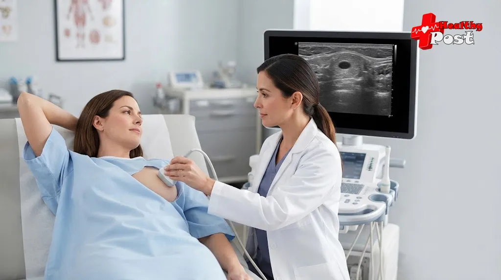
Ultrasound uses sound waves to create images of breast tissue. One of its useful roles is helping distinguish a fluid-filled lesion, such as a cyst, from a solid mass. It can also characterize the shape, margins, orientation, and other features of a breast mass.
For example, a report might describe a nodule as:
- Oval or round
- Circumscribed or irregular
- Solid or fluid-filled
- Parallel or non-parallel to the skin
- Hypoechoic or otherwise different in echogenicity from surrounding tissue
These descriptions are pieces of the radiologist’s assessment. They should not be interpreted individually as proof that a lesion is cancerous.
The most useful part of the report is usually the final assessment and BI-RADS category, along with the recommended next step.
Common Causes of a Breast Nodule
There are several possible explanations for a breast lump or ultrasound finding. Some are completely benign, while others require additional investigation.
Fibroadenoma
A breast fibroadenoma is a common benign solid breast tumor. It often feels smooth, firm, and mobile when it can be felt through the skin. Fibroadenomas are particularly common in younger women and may change in size over time.
On ultrasound, a typical fibroadenoma may have a smooth, oval appearance with well-defined margins. However, imaging characteristics must be interpreted by a qualified clinician rather than used for self-diagnosis.
Some fibroadenomas can simply be monitored. If a lesion grows, develops unusual imaging features, or does not fit the expected appearance, additional assessment or biopsy may be recommended.
Breast cyst
A breast cyst on ultrasound is a fluid-filled structure within the breast. Simple cysts often have characteristic ultrasound features that allow radiologists to recognize them as benign.
Cysts can sometimes become tender, particularly around hormonal changes associated with the menstrual cycle. A painful or large cyst may sometimes be drained with a needle for symptom relief.
Ultrasound is especially useful because it can help determine whether a palpable lump is fluid-filled or solid.
Fibrocystic or hormonal breast changes
Hormonal fluctuations can make breast tissue feel lumpier, fuller, or more tender at certain times of the menstrual cycle. These changes are common and can sometimes produce areas that feel like nodules.
This is one reason the clinical history matters. A lump that changes predictably with the menstrual cycle may have a different evaluation pathway from a new, persistent mass.
Inflammation or infection
Mastitis and breast abscesses can produce a lump or localized area of abnormality. They may be accompanied by pain, warmth, redness, swelling, or fever.
Inflammatory breast findings therefore need to be interpreted alongside symptoms rather than treated as an isolated ultrasound abnormality.
Other benign masses
Other causes can include lipomas, intraductal papillomas, fat necrosis after injury or surgery, and other benign breast conditions. The National Cancer Institute notes that benign breast conditions are common and that not all breast abnormalities increase future breast cancer risk.
For broader context about how breast characteristics and cancer risk differ, you can also read this explanation of breast density and cancer risk. It is important to remember that physical breast size itself is not the same thing as mammographic breast density or cancer risk.
When Could a Breast Nodule Be Suspicious?
Some breast imaging findings require closer evaluation because their appearance is not clearly benign.
Doctors may pay attention to features such as:
- Irregular or indistinct margins
- An unusual orientation within the breast
- Architectural distortion
- Associated abnormalities in surrounding tissue
- Changes compared with earlier imaging
- A mass that is clinically concerning or continues to enlarge
A suspicious appearance does not automatically establish cancer. It means the imaging features are concerning enough that further investigation may be appropriate.
Similarly, symptoms cannot reliably distinguish benign from malignant disease. Breast cancer can cause a lump or changes in breast shape, skin, or the nipple, but early breast cancer may have no obvious symptoms. The NCI emphasizes that many breast changes are not cancer, while still recommending medical evaluation of new or unusual changes.
Breast Cancer Symptoms to Know
Potential breast cancer symptoms can include:
- A new lump in the breast or underarm
- A thick or firm area that is different from surrounding tissue
- A change in breast size or shape
- Skin dimpling or puckering
- Persistent skin changes, redness, or swelling
- A newly inverted nipple
- Unusual nipple discharge, particularly when it is not breast milk
- Persistent changes that do not resolve
However, none of these findings alone proves cancer. Conversely, the absence of symptoms does not completely rule it out because early breast cancer may be clinically silent.
If you notice a new breast lump or a persistent change, arrange an assessment rather than trying to determine its cause from symptoms alone.
Understanding BI-RADS Breast Ultrasound Categories
One of the most important terms on a breast imaging report is BI-RADS.
BI-RADS stands for Breast Imaging Reporting and Data System. The American College of Radiology uses this standardized system for breast imaging, including ultrasound, mammography, and MRI. Its purpose is to give radiologists a consistent vocabulary and connect imaging findings with management recommendations.
Here is a practical overview of the BI-RADS breast classification:
| BI-RADS | General meaning | Typical management |
|---|---|---|
| 0 | Incomplete assessment | Additional imaging or comparison with previous studies |
| 1 | Negative | Routine screening or care as appropriate |
| 2 | Benign finding | Routine screening/follow-up as appropriate |
| 3 | Probably benign | Short-interval imaging follow-up |
| 4 | Suspicious abnormality | Biopsy should be considered |
| 5 | Highly suggestive of malignancy | Appropriate action, generally including tissue diagnosis |
| 6 | Known biopsy-proven malignancy | Management of the confirmed malignancy |
The ACR’s current reporting guidance specifically describes BI-RADS 3 as a probably benign finding for which short-interval follow-up is suggested, BI-RADS 4 as a suspicious abnormality for which biopsy should be considered, and BI-RADS 5 as highly suggestive of malignancy. BI-RADS 6 is reserved for a known biopsy-proven malignancy.
BI-RADS 0 deserves special attention because it does not mean cancer. It means the assessment is incomplete and additional imaging evaluation is needed before a final category can be assigned.
What Does BI-RADS 3 Mean?
BI-RADS 3 means probably benign. It is not the same as a cancer diagnosis.
The BI-RADS framework places the expected likelihood of malignancy for category 3 at no more than 2%, and the usual approach is imaging surveillance rather than immediate biopsy when the finding meets the criteria for this category.
The purpose of short-term follow-up is to make sure the finding remains stable.
A patient should therefore avoid interpreting “follow-up recommended” as “the doctor thinks I have cancer.” It generally means the imaging appearance is sufficiently reassuring to monitor but not appropriate to simply dismiss.
What Does BI-RADS 4 Mean?
BI-RADS 4 means the finding is suspicious and tissue diagnosis should be considered.
Category 4 can be divided into 4A, 4B, and 4C, representing increasing levels of suspicion. Importantly, BI-RADS 4 covers a broad range rather than representing one single probability.
This is why simply seeing “BI-RADS 4” on a report does not tell you exactly what the final diagnosis will be.
What Does BI-RADS 5 Mean?
BI-RADS 5 means the imaging appearance is highly suggestive of malignancy. The ACR describes category 5 as having a very high probability of malignancy, with tissue diagnosis generally required to establish the diagnosis.
Even at this stage, the BI-RADS category is an imaging assessment—not the same thing as a pathology result.
What Does BI-RADS 6 Mean?
BI-RADS 6 is different from categories 4 and 5. It is used when there is already a known biopsy-proven malignancy and imaging is being performed in the context of that confirmed diagnosis.
How Is a Breast Nodule Diagnosed?
A breast nodule diagnosis usually involves several pieces of information rather than one test.
Your healthcare professional may consider:
- Medical and family history
- Clinical breast examination
- Ultrasound
- Diagnostic mammography when appropriate
- MRI in selected situations
- Biopsy when imaging or clinical findings warrant tissue diagnosis
The choice depends on age, symptoms, examination findings, previous imaging, risk factors, and the characteristics of the lesion.
Ultrasound is especially useful for evaluating a palpable lump and characterizing whether a lesion is fluid-filled or solid. Mammography and MRI may provide additional information in selected circumstances.
Why a Breast Nodule Biopsy May Be Recommended
Imaging can sometimes provide enough information to classify a lesion as benign. But when an abnormality is suspicious or cannot be confidently characterized, a breast nodule biopsy may be needed.
A biopsy removes cells or tissue so that a pathologist can examine them under a microscope. NCI describes biopsy as the only sure way to diagnose breast cancer.
Common approaches include:
- Fine-needle aspiration: removes fluid or cells using a thin needle.
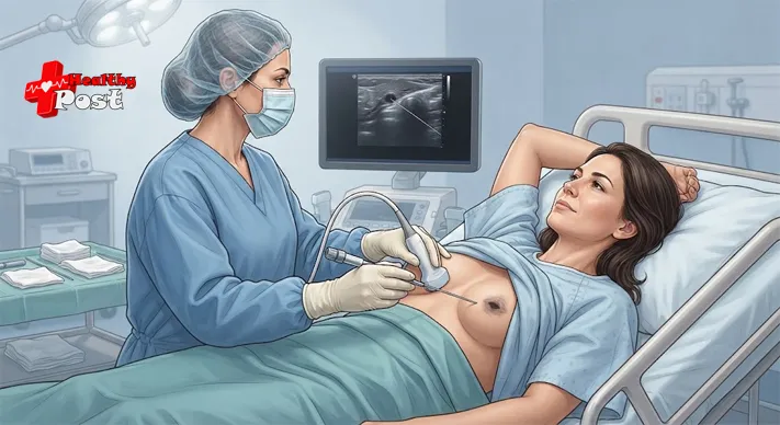
- Core-needle biopsy: removes small cylinders of tissue and is commonly used for suspicious solid masses.
- Image-guided biopsy: ultrasound, mammography, or MRI can help guide the needle precisely.
- Surgical biopsy: removes part or all of a lesion when necessary.
The procedure selected depends on the type and location of the abnormality and the information doctors need.
What Happens After a Breast Nodule Is Found?
The next step depends on the complete assessment.
If the finding is clearly benign
A BI-RADS 2 finding generally does not require biopsy simply because a nodule is present. Routine breast care or screening may be recommended.
For some benign conditions, treatment is considered only if symptoms are significant. For example, a painful cyst may sometimes be drained.
If the finding is probably benign
A BI-RADS 3 finding commonly leads to short-term imaging follow-up. The goal is to document stability rather than immediately perform an invasive procedure.
Keep the recommended appointment even if the lump feels unchanged. Follow-up imaging is part of the diagnostic process.
If the finding is suspicious
A BI-RADS 4 or 5 assessment generally requires further diagnostic evaluation, often including biopsy. The exact procedure depends on the lesion and the imaging modality that best demonstrates it.
If cancer is confirmed
Once pathology confirms breast cancer, treatment is based on factors such as the cancer type, stage, receptor status, and other clinical characteristics. Treatment may involve surgery, radiation therapy, chemotherapy, endocrine therapy, targeted therapy, immunotherapy, or combinations of these depending on the individual diagnosis.
This is a very different stage of care from simply finding a nodule on ultrasound.
For readers trying to understand what a biopsy can reveal about a breast lesion, HealthyPost also has an article discussing atypical ductal hyperplasia and core-needle biopsy findings. That condition is distinct from a simple ultrasound nodule, but the article provides additional context about why tissue sampling can sometimes lead to further diagnostic decisions.
When Should You Seek Medical Assessment?
A new breast lump should not be ignored simply because it is painless or because a previous imaging study was normal.
Arrange medical evaluation if you notice:
- A new or persistent breast lump
- A lump that becomes larger or changes
- A firm or fixed area
- New nipple inversion
- Bloody or unusual nipple discharge
- Skin dimpling or puckering
- Persistent redness or swelling
- A new lump in the armpit
- Breast changes that do not resolve
These signs do not automatically mean cancer. They indicate that the change deserves proper assessment. The NCI specifically advises people to follow up with a healthcare professional when they notice a breast lump or other unusual breast change, even after a recent normal mammogram.
FAQs About Breast Nodules on Ultrasound
Does a breast nodule on ultrasound mean cancer?
No. A breast nodule is a descriptive imaging finding, not a cancer diagnosis. Cysts, fibroadenomas, hormonal changes, inflammation, and other benign conditions can produce breast lumps or nodules. The imaging assessment and, when necessary, biopsy determine what the lesion represents.
Is a solid breast nodule always cancerous?
No. A solid lesion can be benign, including a fibroadenoma. However, some solid masses are malignant, which is why the lesion’s complete ultrasound appearance and BI-RADS assessment matter.
Does BI-RADS 3 mean I have breast cancer?
No. BI-RADS 3 means probably benign and generally leads to short-interval imaging follow-up. It is not a diagnosis of cancer.
Does every breast nodule need a biopsy?
No. Some nodules have imaging features that are sufficiently characteristic of a benign condition that biopsy is not necessary. Suspicious or indeterminate findings may require tissue sampling.
Can a breast cyst become cancer?
A simple breast cyst is a benign fluid-filled lesion and is not itself breast cancer. However, not every breast lump is a simple cyst, so a new or persistent lump should be properly assessed rather than assumed to be a cyst.
What should I do if my ultrasound report says BI-RADS 4?
Arrange the recommended follow-up with your healthcare professional or breast-imaging team. BI-RADS 4 means the finding is suspicious and biopsy should be considered, but the category alone does not establish that cancer is present.
Conclusion
A breast nodule on ultrasound is a finding that needs context, not an automatic cancer diagnosis. Many nodules are caused by benign conditions such as fibroadenomas, cysts, hormonal breast changes, or inflammation.
The most useful information on an imaging report is usually the BI-RADS assessment and management recommendation. BI-RADS 1 and 2 generally indicate negative or benign findings, BI-RADS 3 generally calls for short-term imaging surveillance, while BI-RADS 4 and 5 indicate increasing levels of imaging concern and commonly lead to tissue diagnosis. BI-RADS 6 is reserved for a malignancy that has already been confirmed by biopsy.
If a breast nodule is found, the practical next step is to review the complete report with a qualified healthcare professional. Bring previous imaging if available, ask what the BI-RADS category means, and follow the recommended surveillance or diagnostic plan.
The combination of imaging findings, clinical examination, medical history, and—when necessary—biopsy results provides a much more reliable answer than the word “nodule” alone.
References
- American College of Radiology — BI-RADS®: Breast Imaging Reporting and Data System. The ACR’s current BI-RADS resource explains standardized breast imaging terminology, assessment categories, and management recommendations.
American College of Radiology — BI-RADS® - National Cancer Institute — Signs and Symptoms of Breast Cancer. Provides current information on breast changes, potential symptoms, and the importance of medical follow-up.
National Cancer Institute — Breast Cancer Signs and Symptoms - National Cancer Institute — How Is Breast Cancer Diagnosed? Explains clinical assessment, breast imaging, biopsy, and how tissue diagnosis is established.
National Cancer Institute — How Is Breast Cancer Diagnosed?