
Know everything about Robotic myomectomy
Robotic myomectomy is an advanced gynecologic surgery that offers a sophisticated solution for women dealing with uterine fibroids. This innovative procedure combines robotic technology with minimally invasive techniques to remove non-cancerous tumors from the uterus.
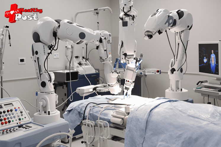
The procedure uses advanced robotic systems, allowing surgeons to perform complex movements through small incisions in the abdomen. A high-definition 3D camera provides clear visualization, while robotic arms accurately replicate the surgeon’s hand movements.
Key advantages of robotic myomectomy include:
- Smaller incisions leading to reduced scarring
- Less blood loss during surgery
- Shorter hospital stays
- Faster recovery times
- Enhanced surgical precision
- Reduced post-operative pain
This groundbreaking approach has transformed fibroid treatment, providing an alternative to traditional open surgery. The technology enables surgeons to preserve the uterus while effectively treating fibroid symptoms, making it particularly valuable for women who wish to maintain their fertility options.
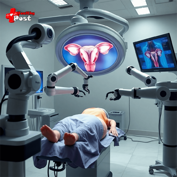
Understanding Uterine Fibroids
Uterine fibroids are non-cancerous tumors that develop within the uterine walls. These growths can vary significantly in size, from as small as a pea to as large as a melon. Medical professionals classify fibroids into distinct types based on their location:
- Intramural fibroids: Grow within the muscular uterine wall
- Subserosal fibroids: Develop on the outer surface of the uterus
- Submucosal fibroids: Project into the uterine cavity
Women with uterine fibroids often experience a range of disruptive symptoms:
- Heavy menstrual bleeding leading to anemia
- Intense pelvic pressure and pain
- Frequent urination
- Lower back pain
- Pain during intercourse
- Difficulty getting pregnant
These symptoms can significantly impact daily life, causing women to miss work, avoid social activities, and experience emotional distress. The physical toll of fibroids extends beyond the immediate symptoms – many women report feeling constantly fatigued due to heavy blood loss and chronic pain.
The presence of fibroids can also affect reproductive health, potentially interfering with conception and pregnancy. Some women may experience pregnancy complications or fertility challenges due to the location and size of their fibroids.
The Robotic Myomectomy Procedure
Robotic myomectomy combines advanced surgical technology with traditional laparoscopic techniques to remove uterine fibroids. This innovative procedure uses the da Vinci Surgical System, featuring robotic arms equipped with specialized surgical instruments and a high-definition 3D camera.
How the Procedure Works:
- The surgeon operates from a dedicated console near the operating table
- Advanced computer technology translates the surgeon’s hand movements into precise micro-movements
- Robotic arms perform the actual surgical work through small abdominal incisions
- A high-definition camera provides detailed 3D visualization of the surgical site
Key Technical Aspects:
- Small Incisions: 3-4 tiny cuts (8-12mm) in the abdomen
- Port Placement: Strategic positioning of specialized tubes for instrument access
- Robotic Docking: Careful attachment of robotic arms to the surgical ports
- Console Control: Surgeon manipulation of controls while viewing 3D imagery
Differences from Traditional Approaches:
| Traditional Laparoscopy Robotic Myomectomy Open Surgery Manual instrument control | Enhanced surgical precision | Large abdominal incision |
| 2D visualization | Superior 3D visualization | Direct manual access |
| Limited range of motion | Greater range of instrument motion | Extended recovery time |
| Longer learning curve | Improved ergonomics for surgeons | Higher risk of complications |
The robotic system filters out natural hand tremors and scales down the surgeon’s movements, enabling exceptional precision during critical steps like fibroid removal and uterine repair. This technology allows surgeons to perform complex surgical maneuvers through tiny incisions with unprecedented control and accuracy.
The robotic platform’s advanced features help surgeons navigate challenging cases, particularly when dealing with multiple fibroids or those in difficult-to-reach locations. The system’s articulating instruments provide enhanced mobility compared to traditional straight-stick laparoscopic tools, allowing for more precise suturing and tissue manipulation.
Patient Selection and Indications for Robotic Myomectomy
Selecting suitable candidates for robotic myomectomy requires careful evaluation of specific criteria to ensure optimal surgical outcomes.
Ideal Candidates:
- Women with symptomatic fibroids who wish to preserve fertility
- Patients with 1-4 fibroids measuring less than 10 cm
- Cases involving intramural or subserosal fibroids
- Individuals with no significant prior abdominal surgeries
- Patients with a BMI under 35
Key Selection Factors:
- Fibroid Location: Subserosal and intramural fibroids are most accessible through robotic approaches
- Size Considerations: Fibroids between 3-8 cm respond best to robotic removal
- Number of Fibroids: Multiple fibroids can increase surgical complexity
- Patient Health Status: Good candidates maintain stable general health
Medical Considerations:
- Previous response to medical management
- Presence of severe symptoms affecting quality of life
- Desire for future pregnancies
- Absence of suspected malignancy
- Normal cervical cytology
The choice of robotic myomectomy often depends on surgeon expertise, hospital resources, and patient-specific anatomical factors. Your surgeon will evaluate imaging studies, medical history, and reproductive goals to determine if robotic myomectomy presents the most appropriate surgical approach for your specific case.
Surgical Steps Involved in Robotic Myomectomy
The surgical process begins with essential preoperative preparations. You’ll need to fast for 8-12 hours before the procedure. Your surgical team will administer general anesthesia to ensure you remain comfortable throughout the operation.
Pre-Surgery Protocol:
- Complete blood work and imaging studies
- Stop certain medications as advised by your surgeon
- Shower with antiseptic soap the night before
- Arrive at the hospital wearing loose, comfortable clothing
The actual surgical procedure follows a systematic approach:
1. Initial Setup
- Creation of 3-4 small incisions (0.5-1 cm) in your lower abdomen
- Insertion of a camera port through the belly button
- Placement of additional ports for robotic arms and surgical instruments
2. Docking Phase
- Positioning of the robotic system next to the operating table
- Connection of robotic arms to the inserted ports
- Surgeon moves to the control console
3. Fibroid Removal Process
- Injection of vasoconstrictive solution to minimize bleeding
- Precise incision of the uterine wall using robotic instruments
- Careful separation of fibroid tissue from healthy uterine muscle
- Extraction of fibroids using specialized grasping tools
4. Repair and Closure
- Multi-layer suturing of the uterine wall
- Meticulous reconstruction to maintain uterine integrity
- Removal of all surgical instruments
- Closure of abdominal incisions with dissolvable stitches
The surgeon maintains complete control throughout the procedure, utilizing the robotic system’s enhanced visualization and precision. The 3D high-definition camera provides detailed views of the surgical field, allowing for accurate identification of tissue planes and blood vessels.
A typical robotic myomectomy takes 2-4 hours, depending on the number, size, and location of fibroids. The hybrid technique combines conventional laparoscopic instruments with robotic technology when needed for optimal surgical outcomes.
Benefits of Robotic Myomectomy Compared to Other Techniques
Robotic myomectomy offers distinct advantages that set it apart from traditional surgical approaches. The da Vinci robotic system provides surgeons with enhanced capabilities:
1. Superior 3D Visualization
- High-definition magnified views of the surgical field
- Clear depth perception for precise tissue handling
- Better identification of anatomical structures
2. Enhanced Surgical Precision
- Robotic arms filter out natural hand tremors
- 360-degree instrument rotation
- Scaled movements for delicate procedures
The robotic platform transforms how surgeons perform complex procedures:
3. Improved Dexterity
- Wristed instruments mimic human hand movements
- Better access to hard-to-reach areas
- Precise suturing in confined spaces
4. Patient Benefits
- Smaller incisions compared to open surgery
- Reduced blood loss during the procedure
- Shorter hospital stays
- Faster return to daily activities
- Minimal scarring
The technological advantages of robotic surgery translate into measurable clinical outcomes. Studies show patients experience less post-operative pain and require fewer pain medications compared to traditional open surgery. The precision of robotic instruments allows surgeons to perform complex fibroid removals while preserving healthy uterine tissue.
Risks and Potential Complications Associated with Robotic Myomectomy
Like any surgical procedure, robotic myomectomy carries specific risks you should understand before proceeding with the treatment.
Common Surgical Risks:
- Post-operative infections at incision sites
- Excessive bleeding during or after surgery
- Formation of blood clots in legs or pelvic area
- Adverse reactions to anesthesia
- Development of adhesions (scar tissue)
Procedure-Specific Complications:
- Damage to surrounding organs, including:
- Bladder
- Bowel
- Ureters
- Blood vessels
- Conversion to open surgery if complications arise
- Incomplete removal of fibroids
- Weakening of uterine wall
Post-Surgical Considerations:
- Risk of uterine rupture in future pregnancies
- Possibility of fibroid recurrence
- Extended operative time compared to traditional methods
- Potential mechanical failure of robotic equipment
Your surgeon will implement specific preventive measures to minimize these risks:
- Pre-operative imaging to map fibroid locations
- Careful patient positioning
- Strategic placement of robotic arms
- Continuous monitoring during the procedure
- Meticulous closure of the uterine wall
The risk of complications varies based on:
- Size and location of fibroids
- Number of fibroids being removed
- Your overall health status
- Previous abdominal surgeries
- Surgeon’s experience with robotic procedures
Studies show complication rates for robotic myomectomy remain relatively low when performed by experienced surgeons in properly selected patients.
Recovery After Robotic Myomectomy Surgery
The recovery journey after robotic myomectomy requires careful attention to postoperative care guidelines. You can expect to stay in the hospital for 24-48 hours after surgery, allowing medical staff to monitor your vital signs and manage any immediate discomfort.
Initial Recovery Phase (First Week)
During the first week after surgery, you can expect the following:
- Light vaginal bleeding or spotting lasting 5-7 days
- Prescribed pain medications to manage discomfort
- Rest and limited movement around your home
- Small sips of water and gradual return to normal diet
Activity Restrictions (2-4 Weeks)
In the 2-4 weeks following surgery, it is important to adhere to these activity restrictions:
- No heavy lifting (over 10 pounds)
- Avoid strenuous exercise or vigorous activities
- Limited climbing of stairs
- No sexual activity
- No swimming or bathing (showers permitted)
Return to Normal Activities
You can gradually resume normal activities as follows:
- Light household tasks after 1 week
- Desk work or remote work after 1-2 weeks
- Driving once off pain medication (typically 1 week)
- Regular exercise at 4-6 weeks
- Full recovery within 6 weeks
Signs of Normal Healing
Keep an eye out for these signs of normal healing:
- Decreasing abdominal pain
- Minimal incision site drainage
- Reducing vaginal discharge
- Improved energy levels daily
- Gradual increase in mobility
Your surgeon will schedule follow-up appointments at one week and six weeks post-surgery to assess your healing progress. These visits allow for incision checks, discussion of any concerns, and clearance for returning to normal activities.
Impact on Future Pregnancies After Robotic Myomectomy
Pregnancy planning after robotic myomectomy requires careful consideration and medical guidance. Your healthcare provider will discuss specific waiting periods before attempting conception – typically 4-6 months after surgery to allow proper healing of the uterine wall.
Key Pregnancy Considerations:
- The location and depth of fibroid removal impacts uterine wall strength
- Multiple or large fibroids increase potential pregnancy risks
- Previous cesarean sections affect delivery method recommendations
The surgical technique used during robotic myomectomy influences future pregnancy management. Your doctor will document the specific details of your procedure, including:
- Number and size of removed fibroids
- Depth of uterine wall involvement
- Type of sutures used for repair
- Any complications during surgery
Most healthcare providers recommend cesarean delivery for pregnancies following robotic myomectomy. This recommendation stems from concerns about uterine rupture during labor contractions. The risk varies based on:
- Surgical technique used
- Healing quality of the uterine wall
- Number and size of removed fibroids
- Time elapsed since surgery
Regular prenatal monitoring becomes essential during pregnancy after myomectomy. Your healthcare team will track uterine scar integrity through ultrasound assessments and carefully monitor your pregnancy progression.
Cost Considerations and Surgeon Expertise in Robotic Myomectomy
Robotic myomectomy costs typically range from $15,000 to $25,000, significantly higher than traditional laparoscopic procedures. The increased expense stems from:
- Advanced robotic equipment maintenance
- Specialized surgical instruments
- Extended operating room time
- Additional training requirements
Insurance coverage varies by provider and plan type. Many insurance companies classify robotic myomectomy as an elective procedure, potentially leading to higher out-of-pocket expenses.
Surgeon expertise plays a crucial role in successful outcomes. Key factors include:
- Completion of specialized robotic surgery training
- Experience with complex fibroid cases
- Regular performance of robotic procedures
- Understanding of patient selection criteria
The learning curve for surgeons transitioning to robotic platforms can be steep. Research indicates surgeons need to perform 20-30 cases to achieve proficiency. Hospitals often require surgeons to maintain a minimum annual case volume to retain robotic surgery privileges.
You should discuss cost options with your healthcare provider and insurance company to understand coverage details and potential payment plans.
Is Robotic Myomectomy Right for You? Consult with an Expert Surgeon!
Your journey toward treating uterine fibroids starts with a personalized assessment of your specific condition. The decision to undergo robotic myomectomy depends on several key factors:
- Fibroid Characteristics
- Size and location of your fibroids
- Number of fibroids present
- Growth patterns and symptoms
- Personal Health Factors
- Age and medical history
- Future pregnancy plans
- Previous surgical procedures
- Current symptoms and their impact on daily life
The best way to determine if robotic myomectomy suits your needs is through a consultation with an experienced gynecologic surgeon. During your consultation, you’ll:
- Review your complete medical history
- Discuss available treatment alternatives
- Understand the potential risks and benefits
- Get answers to your specific questions
- Create a personalized treatment plan
Schedule an appointment with a qualified surgeon to explore whether robotic myomectomy aligns with your treatment goals. Your surgeon will guide you through the decision-making process, ensuring you make an informed choice about your fibroid treatment journey.
FAQs (Frequently Asked Questions)
What is robotic myomectomy and how does it treat uterine fibroids?
Robotic myomectomy is a minimally invasive surgical procedure that uses robotic technology to precisely remove uterine fibroids, which are non-cancerous tumors. This technique offers enhanced dexterity and 3D visualization for surgeons, making it an effective treatment option for women with symptomatic fibroids.
What types of uterine fibroids can be treated with robotic myomectomy?
Robotic myomectomy is ideal for treating intramural and subserosal fibroids. The suitability depends on the size, number, and location of the fibroids, with patient selection being crucial to achieving optimal outcomes.
How does robotic myomectomy compare to traditional laparoscopic and open surgery methods?
Compared to traditional laparoscopy and open surgery, robotic myomectomy provides greater surgical precision through robotic arms controlled from a console. It reduces tremors, allows smaller incisions, minimizes blood loss, and often results in quicker recovery times due to its minimally invasive nature.
What are the common risks and potential complications associated with robotic myomectomy?
While robotic myomectomy is generally safe, potential risks include infection, excessive bleeding, blood clots, and injury to surrounding organs. These risks are similar to those in other surgical procedures but may be reduced due to the precision of robotic technology.
What should patients expect during recovery after robotic myomectomy surgery?
Postoperative care involves managing vaginal bleeding that may last for some time after surgery and adhering to activity restrictions during the initial recovery phase. Recovery timelines vary, but minimally invasive techniques typically allow for faster healing compared to open surgery.
How does robotic myomectomy impact future pregnancies and delivery methods?
Patients are counseled about pregnancy considerations following robotic myomectomy. While many women can conceive successfully post-surgery, cesarean delivery is often recommended to reduce risks related to uterine integrity after fibroid removal.