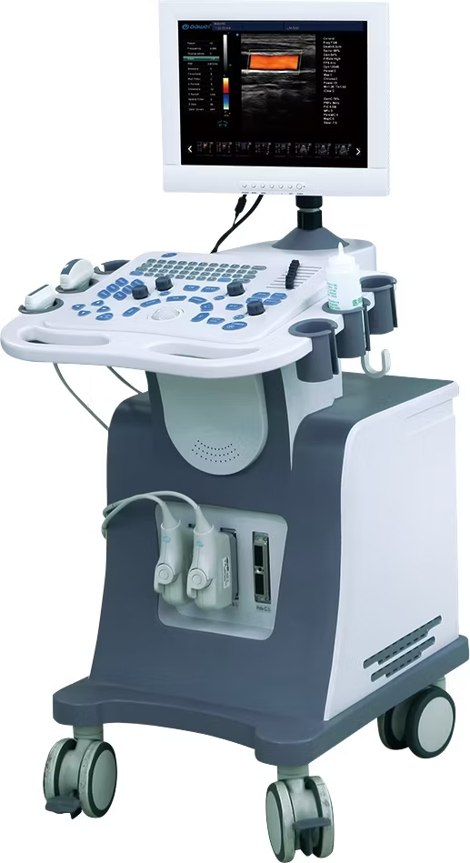
A complete guide to 4D color ultrasound
Origin of the name: Four-dimensional color ultrasound (4D color ultrasound) is also called real-time three-dimensional dynamic image . The so-called “dimension” refers to the spatial dimension. From a scientific point of view, there is only three dimensions, and there is no such thing as four dimensions. “Four-dimensional color ultrasound” actually counts time as one dimension, indicating that three-dimensional images can be obtain in real time. So it seems that “four-dimensional” is just a hype concept.
Function: Can “four-dimensional color ultrasound” really be clearer than other B-ultrasound examinations? Not necessarily. For fetal anomaly screening , the effect of “four-dimensional color ultrasound” is no different from that of ordinary three-dimensional color ultrasound. Therefore, it does not seem to be necessary to do “four-dimensional color ultrasound” specifically for anomaly screening.

Advantages: “ Four-dimensional color ultrasound” can obtain dynamic images of the fetus in the womb, and some hospitals can also burn these images into CDs for expectant mothers to keep. Leaving a memory of the baby’s fetal period may be the main reason to attract expectant mothers.
Expectant mothers talk about their experiences
After the 4D color Doppler ultrasound, the doctor gave me two photos, but no CD. After returning home, I compared the photos taken by my friend with the photos taken by the ordinary 3D color Doppler ultrasound. I found that the photos taken by the two methods were similar. If I had known earlier, I would have done the 3D one. – Xiaoba, 24 weeks pregnant
When doing the 4D color ultrasound, the doctor allowed me to look at the screen, but only showed the baby’s face and limbs, not the key parts that can determine the gender. I wanted to go home and watch the disc to find out more, but I found that the disc didn’t show that part either. Oh… that’s the policy. — Dingdang, 22 weeks pregnant
The hospital I went to for the checkup offere two options: one was to pay the hospital to burn a 4D color Doppler ultrasound disc, and the other was to record the video yourself while it was being done. I chose to record the video myself, but the result was not as clear as the disc burned by my friend. – 27 weeks pregnant
“Four-dimensional Color Doppler Ultrasound” Tips
After reading the above introduction, you must have known something about “four-dimensional color Doppler ultrasound”. After unveiling its “magical” appearance, whether it is necessary to do it once depends on your decision. The following are some precautions for expectant mothers who want to do “four-dimensional color Doppler ultrasound”.
When is the best time to do it? Four-dimensional color Doppler ultrasound can be do at any gestational age. But it should be emphasize that there is still controversy about doing four-dimensional color Doppler ultrasound in early pregnancy (before 12 weeks of pregnancy). For safety reasons, it is recommend to do it after 16 weeks of pregnancy. The best time is 6 to 8 months of pregnancy (26 to 32 weeks), because during this period, the fetus’s limbs and major organs have all developed, and the amount of amniotic fluid is relatively moderate.
Will it affect the health of the fetus? In fact, “four-dimensional color ultrasound” is also a means of ultrasound examination (B-ultrasound), so there must be a small amount of radiation. Experts recommend that you do not do more than three ultrasound examinations during pregnancy, that is, you can choose “four-dimensional color ultrasound” once out of three times. However, it is not recommend to increase the number of B-ultrasounds in order to take so-called “fetal portraits” for the baby. Even in the United States, there is a clear recommendation that pregnant women should not do that kind of “gift-like ultrasound examination.”
Interpretation of 4D Color Doppler Ultrasound Report
Color Doppler ultrasound
Ultrasound findings: single fetus in utero, BPD 73mm, head circumference 263mm, RSA skull halo is still regular, brain midline is center. Lateral ventricle has no obvious expansion, posterior cranial fossa is 8mm, upper lip has no obvious interruption, spine is still regular, ulna and radius can be displayed, both kidneys can be displayed, gastric bubble can be displayed, four-chamber heart can be displayed, foramen ovale is 5mm with valve, fetal heart beats regularly, 145 times/min, tibia and fibula can be displayed, femur is about 52mm long, placenta is located on the posterior wall of the uterine body, parenchyma echo is uniform, amniotic fluid is about 65mm, and internal sound transmission is good. U-shaped pressure mark is see on the fetal neck, CDFI: umbilical cord blood flow signal is around the neck. Placental function: Grade Ι Ultrasound prompts : single fetus breech presentation, about 29 weeks, umbilical cord around the neck
Color Doppler ultrasound term analysis:
BPD: Biparietal diameter, which refers to the distance between the two parietal bones of the fetus. Doctors often use it to observe the development of the child, determine whether there is cephalopelvic disproportion, and smoothly deliver the baby . Head circumference: The length of the circumference of the head. RSA:
The fetal position is breech. Femoral length (also call FL): The femur is the largest long bone in the human body, referring to the thigh bone. It is a common indicator use by doctors to observe the development of the fetus when they use B-ultrasound to check the pregnant mother during pregnancy. Placental function level: There are one, two, and three levels of placental function. Level one means that the current uterine environment is more suitable for fetal growth. Once it reaches level three, the placenta will calcify and its function will tend to decline.
Umbilical cord around the neck for X weeks: Umbilical cord around the neck is a situation that many pregnant mothers will encounter. Generally speaking, it has little impact. As long as the pregnant mother lies flat more, pats the belly gently, and lets the fetus move more in the belly, some babies can untie the umbilical cord around the neck by themselves.
The umbilical cord is 50 cm long and has no effect on the fetus. Whether the baby will be born naturally is not determined by the umbilical cord around the neck. It should be determined with the obstetrician and gynecologist before delivery. Occipital-frontal diameter: the distance from the root of the fetal nose to the occipital protuberance. Abdominal circumference: the length of the abdomen. No obvious interruption of the upper lip: no cleft lip. Regular arrangement of the spine: normal spine, kidney, stomach, heart. Tibia and fibula: lower leg.

